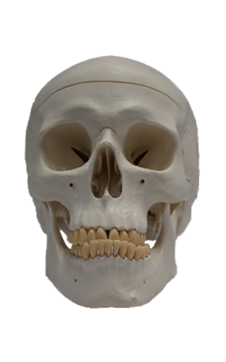
Main Model

BRAIN : Right lateral view of right brain

Brain
Because the brain is usually studied in detail in a separate neuroanatomy course, the brain is covered by only a
superficial survey of its gross structure in the typical anatomy course, with attention primarily concerned with the
relationship between the brain and its environment - that
is, its meningeal coverings, the CSF-filled subarachnoid
space, and internal features of its bony encasement (the
neurocranium).
Because of their role in the production of CSF (cerebrospinal fluid), the ventricles of the brain and the CSF-producing
choroid plexuses found there are also covered. Furthermore,
11 of 12 cranial nerves arise from the brain.
Parts of Brain
The brain (contained by the neurocranium) is composed
of the cerebrum, cerebellum, and brainstem.
When the calvaria and dura are removed, gyri (folds), sulci (grooves), and fissures (clefts) of the cerebral cortex are visible through the delicate arachnoid-pia layer. Whereas the gyri and sulci demonstrate much variation, the other features
of the brain, including overall brain size, are remarkably consistent from individual to individual.
• The cerebrum (Latin brain) includes the cerebral hemispheres and basal ganglia. The cerebral hemispheres,
separated by the falx cerebri within the longitudinal
cerebral fissure, are the dominant features of the brain. Each cerebral hemisphere is divided
for descriptive purposes into four lobes, each of which
is related to, but the boundaries of which do not correspond to, the overlying bones of the same name. From
a superior view, the cerebrum is essentially divided into
quarters by the median longitudinal cerebral fissure and
the coronal central sulcus. The central sulcus separates
the frontal lobes (anteriorly) from the parietal lobes (posteriorly). In a lateral view, these lobes lie superior to
the transverse lateral sulcus and the temporal lobe inferior to it. The posteriorly placed occipital lobes are separated from the parietal and temporal lobes by the plane of
the parieto-occipital sulcus, visible on the medial surface of the cerebrum in a hemisected brain. The anteriormost points of the anteriorly projecting frontal and temporal lobes are the frontal and temporal poles. The posteriormost point of the posteriorly projecting occipital lobe is the occipital pole. The hemispheres
occupy the entire supratentorial cranial cavity. The frontal lobes occupy the anterior cranial fossae,
the temporal lobes occupy the lateral parts of the middle
cranial fossae, and the occipital lobes extend posteriorly
over the tentorium cerebelli.
• The diencephalon is composed of the epithalamus, dorsal thalamus, and hypothalamus and forms the central
core of the brain.
• The midbrain, the rostral part of the brainstem, lies at
the junction of the middle and posterior cranial fossae.
CN III and IV are associated with the midbrain.
• The pons is the part of the brainstem between the midbrain rostrally and the medulla oblongata caudally; it lies
in the anterior part of the posterior cranial fossa. CN V is
associated with the pons.
• The medulla oblongata (medulla) is the most caudal
subdivision of the brainstem that is continuous with the
spinal cord; it lies in the posterior cranial fossa. CN IX,
X, and XII are associated with the medulla, whereas CN
VI-VIII are associated with the junction of pons and medulla.
• The cerebellum is the large brain mass lying posterior to
the pons and medulla and inferior to the posterior part of
the cerebrum. It lies beneath the tentorium cerebelli in the
posterior cranial fossa. It consists of two lateral hemispheres
that are united by a narrow middle part, the vermis.