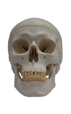
Main Model

CRANIUM : Zygomatic arch

Lateral Aspect of Cranium
The lateral aspect of the cranium is formed by both neurocranium and viscerocranium (Figs. 7.1A & B and 7.4A).
The main features of the neurocranial part are the temporal
fossa, the external acoustic meatus opening, and the mastoid process of the temporal bone. The main features of the
viscero cranial part are the infratemporal fossa, zygomatic
arch, and lateral aspects of the maxilla and mandible.
The temporal fossa is bounded superiorly and posteriorly by the superior and inferior temporal lines, anteriorly by the frontal and zygomatic bones, and inferiorly by the
zygomatic arch (Figs. 7.1A and 7.4A). The superior border of this arch corresponds to the inferior limit of the cerebral
hemisphere of the brain. The zygomatic arch is formed by
the union of the temporal process of the zygomatic bone
and the zygomatic process of the temporal bone.
In the anterior part of the temporal fossa, 3–4 cm superior
to the midpoint of the zygomatic arch, is a clinically important area of bone junctions: the pterion (G. pteron, wing)
(Figs. 7.4A and 7.6; Table 7.1). It is usually indicated by an
H-shaped formation of sutures that unite the frontal, parietal, sphenoid (greater wing), and temporal bones. Less commonly, the frontal and temporal bones articulate; sometimes
all four bones meet at a point.
The external acoustic meatus opening (pore) is the
entrance to the external acoustic meatus (canal), which leads to
the tympanic membrane (eardrum) (Fig. 7.4A). The mastoid
process of the temporal bone is postero-inferior to the external acoustic meatus opening. Anteromedial to the mastoid process is the styloid process of the temporal bone, a slender
needle-like, pointed projection. The infratemporal fossa is an
irregular space inferior and deep to the zygomatic arch, and
the mandible and posterior to the maxilla (see Fig. 7.67B).