Eye, 3 times full size

Admin

Main Model


Orbit : 20 Zygomatic bone

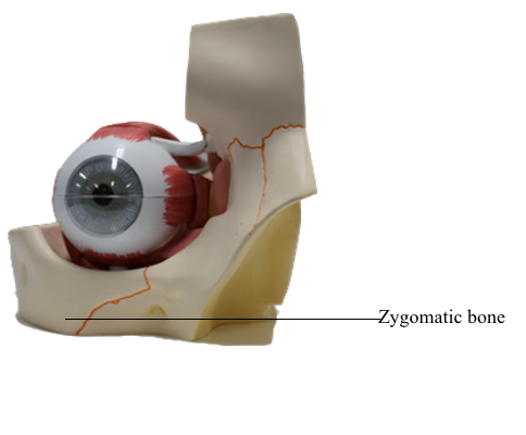
The zygomatic bones (cheek bones, malar bones), forming the prominences of the cheeks, lie on the inferolateral sides of the orbits and rest on the maxillae. The anterolateral rims, walls, floor, and much of the infra-orbital margins of the orbits are formed by these quadrilateral bones. A small zygomaticofacial foramen pierces the lateral aspect of each bone. The zygomatic bones articulate with the frontal, sphenoid, and temporal bones and the maxillae.
  • Extra-ocular muscles
    • I Superior rectus
    • II Inferior rectus
    • III Medial rectus
    • IV Lateral rectus
    • V Tendon of superior oblique 
    • VI Inferior oblique 
  • Fibrous layer of eyeball
    • 1 Cornea
    • 2 Sclera
  • Uvea
    • 3 Iris with pupil
    • 4 Ciliary muscle
    • 5 Corona ciliaris
    • 6 Choroid
    • 7 Ciliary nerves
    • 8 Vorticose vein
    • a Ciliary arteries
  • Inner layer of eyeball
    • 9 Retina
    • 10 Ciliary part of retina
    • 11 Retinal arterioles
    • 12 Retinal venules
    • 13 Macula and fovea centralis
    • 14 Blind spot
    • 15 Lens
    • 16 Vitreous body
  • Orbit
    • 17 Lacrimal sac
    • 18 Optic nerve
    • 19 Optic chiasm
    • 20 Zygomatic bone
    • 21 Maxilla
    • 22 Infra-orbital foramen
    • 23 Nasal bone
    • 24 Frontal bone
    • 25 Frontal sinus
    • 26 Perpendicular plate
    • 27 Crista galli
    • 28 Cribriform plate
    • 29 Spenoidal bone
    • 30 Sphenoidal sinus
    • 31 Superior orbital fissure
    • 32 Foramen rotundum

Copyright 2018, Chulabhorn International College of Medicine

www.cicm.tu.ac.th