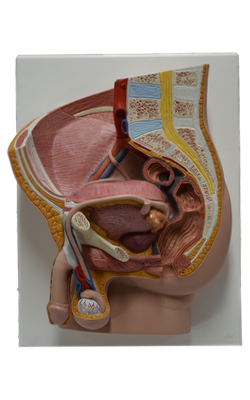
Main Model

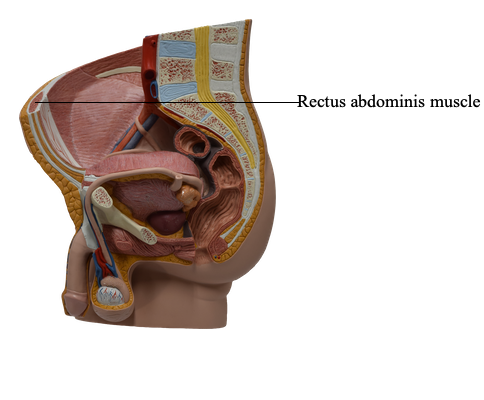
Rectus abdominis muscle

Muscles of Anterolateral Abdominal Wall
There are five (bilaterally paired) muscles in the anterolateral abdominal wall: three flat muscles and two vertical muscles.
The two vertical muscles of the anterolateral abdominal wall, contained within the rectus sheath, are the large rectus abdominis and the small pyramidalis.
Rectus Abdominis Muscle
A long, broad, strap-like muscle, the rectus abdominis (Latin rectus, straight) is the principal vertical muscle of the anterior abdominal wall. The paired rectus muscles, separated by the linea alba, lie close together inferiorly. The rectus abdominis is three times as wide superiorly as inferiorly; it is broad and thin superiorly and narrow and thick inferiorly. Most of the rectus abdominis is enclosed in the rectus sheath. The rectus muscle is anchored transversely by attachment to the anterior layer of the rectus sheath at three or more tendinous intersections. When tensed in muscular people, the areas of muscle between the tendinous intersections bulge outward. The intersections, indicated by grooves in the skin between the muscular bulges, usually occur at the level of the xiphoid process, umbilicus, and halfway between these structures.
Rectus abdominis
Origin: Pubic symphysis and pubic crest
Insertion: Xiphoid process and 5th-7th costal cartilages
Innervation: Thoraco-abdominal nerves (anterior rami of T6-T12 spinal nerves)
Main Action(s): Flexes trunk (lumbar vertebrae) and compresses abdominal viscera; stabilizes and controls tilt of pelvis (antilordosis)