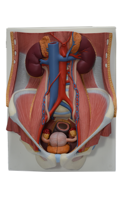
Main Model

5 Abdominal aorta

Most arteries supplying the posterior abdominal wall arise from the abdominal aorta. The subcostal arteries arise from the thoracic aorta and distribute inferior to the 12th rib. The abdominal aorta is approximately 13 cm in length. It begins at the aortic hiatus in the diaphragm at the level of the T12 vertebra and ends at the level of the L4 vertebra by dividing into the right and left common iliac arteries. The abdominal aorta may be represented on the anterior abdominal wall by a band (approximately 2 cm wide) extending from a median point, approximately 2.5 cm superior to the transpyloric plane to a point slightly (2-3 cm) inferior to and to the left of the umbilicus at the level of the supracristal plane (plane of the highest points of the iliac crests). In children and lean adults, the lower abdominal aorta is sufficiently close to the anterior abdominal wall that its pulsations may be detected or apparent when the wall is relaxed.
The common iliac arteries diverge and run inferolaterally, following the medial border of the psoas muscles to the pelvic brim. Here each common iliac artery divides into the internal and external iliac arteries. The internal iliac artery enters the pelvis. The external iliac artery follows the iliopsoas muscle. Just before leaving the abdomen, the external iliac artery gives rise to the inferior epigastric and deep circumflex iliac arteries, which supply the anterolateral abdominal wall.
Relations of Abdominal Aorta
From superior to inferior, the important anterior relations of the abdominal aorta are the:
• Celiac plexus and ganglion.
• Body of the pancreas and splenic vein.
• Left renal vein.
• Horizontal part of the duodenum.
• Coils of small intestine.
The abdominal aorta descends anterior to the bodies of the T12-L4 vertebrae. The left lumbar veins pass posterior to the aorta to reach the inferior vena cava (IVC). On the right, the aorta is related to the azygos vein, cisterna chyli, thoracic duct, right crus of the diaphragm, and right celiac ganglion. On the left, the aorta is related to the left crus of the diaphragm and the left celiac ganglion.
Branches of the Abdominal Aorta
The branches of the descending (thoracic and abdominal) aorta may be described as arising and coursing in three “vascular planes” and can be classified as being visceral or parietal and paired or unpaired. Paired parietal branches of the aorta serve the diaphragm and posterior abdominal wall.
The median sacral artery, an unpaired parietal branch, may be said to occupy a fourth (posterior) plane because it arises from the posterior aspect of the aorta just proximal to its bifurcation. Although markedly smaller, it could also be considered a midline “continuation” of the aorta, in which case its lateral branches, the small lumbar arteries and lateral sacral branches, would also be included as part of the paired parietal branches.