Eye 5, times full size, 8 part

Admin

Main Model


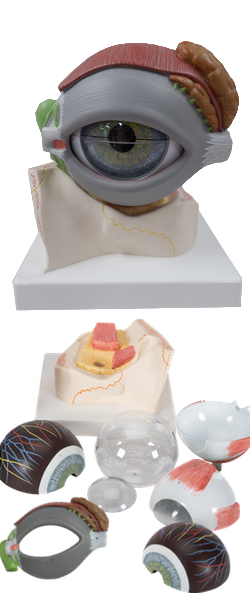
INNER LAYER OF EYEBALL : 12 Retinal venules

Retinal venules (wider) and retinal arterioles (narrower) radiate from the center of the oval optic disc. The dark area lateral to the disc is the macula. Branches of retinal vessels extend toward this area, but do not reach its center, the fovea centralis - the area of most acute vision.
  • EXTRA-OCULA MUSCLES
    • I Superior rectus
    • II Inferior rectus
    • III Medial rectus
    • IV Lateral rectus
    • V Tendon of superior oblique
    • VI Inferior oblique
  • FIBROUS LAYER OF EYEBALL
    • 1 Cornea
    • 2 Sclera
  • VASCULAR LAYER OF EYEBALL
    • 3 Iris with pupil
    • 4 Ciliary muscle
    • 5 Corona ciliaris
    • 6 Choroid
    • 7 Ciliary nerves
    • 8 Vorticose vein
    • 8a Ciliary arteries
  • INNER LAYER OF EYEBALL
    • 9 Retina
    • 10 Ciliary part of retina
    • 11 Retinal arterioles
    • 12 Retinal venules
    • 13 Macula and fovea centralis
    • 14 Optic disc
    • 15 Lens
    • 16 Vitreous body
  • EYELID
    • 1 Levator palpebrae superioris
    • 2 Superior tarsus
    • 3 Inferior tarsus
    • 4 Medial palpebral ligament
    • 5 Lateral palpebral ligament
    • 6 Superior conjunctinal fornix
    • 7 Palpebral conjunctiva
    • 8 Tarsal glands
  • LACRIMAL APPARATUS
    • 9 Lacrimal gland
    • 9a Orbital part
    • 9b Palpebral part
    • 9c Excretory ducts
    • 10 Lacrimal sac
    • 11 Lacus lacrimalis
    • a Superior lacrimal canaliculus
    • b Inferior lacrimal canaliculus
    • c Lacrimal caruncle
    • 12 Lacrimal papilla, lacrimal punctum

Copyright 2018, Chulabhorn International College of Medicine

www.cicm.tu.ac.th