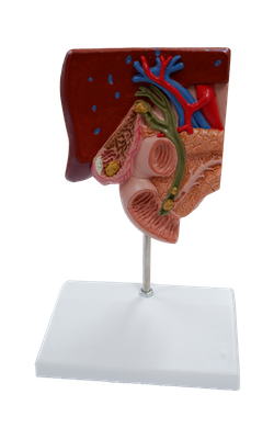
Gallstone Model

Admin

Main Model


Right hepatic duct

Bile, produced by the right and left livers, passes from the liver via the biliary ducts - right hepatic duct and left hepatic duct - that join to form the common hepatic duct, which unites with the cystic duct to form the (common) bile duct and then, conveys all bile from the liver to the descending part of the duodenum.
  • Liver
  • Left hepatic duct
  • Right hepatic duct
  • Common hepatic duct
  • Gallbladder
  • Neck of gallbladder
  • Body of gallbladder
  • Fundus of gallbladder
  • Mucosal fold
  • Spiral fold
  • Cystic duct
  • Common bile duct
  • Major duodenal papilla, major caruncle of Vater
  • Duodenum
  • Sphincter ampullae muscle
  • Abdominal aorta
  • Portal vein of liver
  • Pancreatic duct
  •  Pancreas
  • Gallstone in the fundus area of the gallbladder
  • Gallstone in the area of the spiral valve
  • Wall area with acute inflammation of the gallbladder wall (cholecystitis)
  • Wall area following chronic inflammation of the gallbladder wall: scarred, contracted and thickened
  • Gallstone in the area of the common bile duct
  •  A stone obstructing the papillary opening to the small intestine. Consequently, drainage of the common bile duct and the pancreatic duct is obstructed.

Copyright 2018, Chulabhorn International College of Medicine

www.cicm.tu.ac.th