Lung Model with larynx, 7 part

Admin

Main Model


C TRACHEA : 3 Right subclavain vein

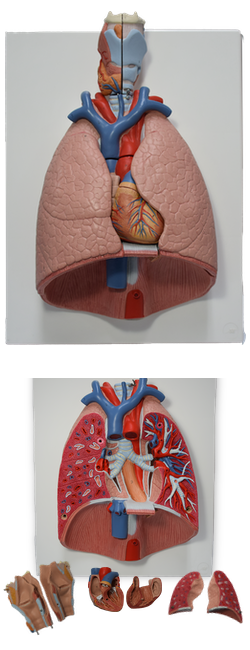
Vein in Root of Neck
The subclavian vein, the continuation of the axillary vein, begins at the lateral border of the 1st rib and ends when it unites with the IJV. The subclavian vein passes over the 1st rib anterior to the scalene tubercle parallel to the subclavian artery, but it is separated from it by the anterior scalene muscle. It usually has only one named tributary, the EJV.
  • 1 Hyoid bone
  • B THYROID GLAND
  • D ESOPHAGUS
  • G DIAPHRAGM
  • A LARYNX
    • 2 Thyroid cartilage
    • 3 Cricoid cartilage
    • 4 Epiglottis
    • 5 Arytenoid carilage
    • 6 Thyrohyoid membrane
    • 7 Thyrohyoid
    • 8 Cricothyroid
    • 9 Transverse arytenoid
    • 10 Oblique arytenoid
    • 11 Posterior crico-arytenoid
    • 12 Laryngeal ventricle
    • 13 Superior laryngeal artery
    • 14 Inferior thyroid vein
  • C TRACHEA
    • 1 Tracheal bifurcation
    • 2 Right main bronchus
    • 3 Right subclavain vein
    • 3a Right subclavian artery
    • 4 Right common carotid artery
    • 5 Right internal jugular vein
    • 6 Right brachiocephalic vein
    • 7 Brachiocephalic trunk
    • 8 Pulmonary artery
  • E LUNG
    • Right lung
    • a Superior lobe
    • b Middle lobe
    • c Inferior lobe
    • Left lung
    • d Superior lobe
    • e Inferior lobe
  • F HEART
    • 9 Left ventricle
    • 10 Ascending aorta
    • 11 Aortic valve
    • 13 Left atrium
    • 14 Left auricle
    • 15 Mitral valve [Left atrioventricular valve]
    • 16 Right ventricle
    • 17 Pulmonary trunk
    • 18 Pulmonary valve
    • 19 Right atrium
    • 20 Right auricle
    • 21 Superior vena cava
    • 22 Inferior vena cava
    • 23 Tricuspid valve [Right atrioventricular valve]
    • 24 Pulmonary veins

Copyright 2018, Chulabhorn International College of Medicine

www.cicm.tu.ac.th