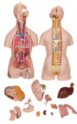
Main Model

ARTERIES

The right common carotid artery begins at the bifurcation of the brachiocephalic trunk. The right subclavian
artery is the other branch of this trunk. From the arch of
the aorta, the left common carotid artery ascends into the
neck. Consequently, the left common carotid has a course
of approximately 2 cm in the superior mediastinum before
entering the neck.
---------------------------------------------------------------------------------------------------------------------------------------------------------------
The subclavian arteries supply the upper limbs; they
also send branches to the neck and brain (Figs. 8.19 and
8.24). The right subclavian artery arises from the brachiocephalic trunk. The left subclavian artery arises from
the arch of the aorta, about 1 cm distal to the left common
carotid artery.
---------------------------------------------------------------------------------------------------------------------------------------------------------------
The brachiocephalic trunk, the fi rst and largest branch
of the arch of the aorta, arises posterior to the manubrium,
where it is anterior to the trachea and posterior to the left
brachiocephalic vein (Figs. 1.65A & B, 1.66B, and 1.68A).
The trunk ascends superolaterally to reach the right side of
the trachea and the right SC joint, where it divides into the
right common carotid and right subclavian arteries.
---------------------------------------------------------------------------------------------------------------------------------------------------------------
The arch of the aorta (aortic arch), the curved continuation of the ascending aorta (Figs. 1.65A and 1.67; see Table
1.5), begins posterior to the 2nd right sternocostal (SC) joint
at the level of the sternal angle. It arches superiorly, posteriorly and to the left, and then inferiorly. The arch ascends
anterior to the right pulmonary artery and the bifurcation of
the trachea, reaching its apex at the left side of the trachea and esophagus as it passes over the root of the left lung. The arch
descends posterior to the left root of the lung beside the T4
vertebra. The arch ends by becoming the thoracic (descending) aorta posterior to the 2nd left sternocostal joint.
---------------------------------------------------------------------------------------------------------------------------------------------------------------
The ascending aorta, approximately 2.5 cm in diameter,
begins at the aortic orifi ce. Its only branches are the coronary
arteries, arising from the aortic sinuses (Fig. 1.55B). The
ascending aorta is intrapericardial (Figs. 1.66A & B); for this
reason, and because it lies inferior to the transverse thoracic
plane, it is considered a content of the middle mediastinum
(part of inferior mediastinum).
---------------------------------------------------------------------------------------------------------------------------------------------------------------
ABDOMINAL AORTA
Most arteries supplying the posterior abdominal wall arise
from the abdominal aorta (Fig. 2.98A; Table 2.15). The subcostal arteries arise from the thoracic aorta and distribute
inferior to the 12th rib. The abdominal aorta is approximately 13 cm in length. It begins at the aortic hiatus in the
diaphragm at the level of the T12 vertebra and ends at the
level of the L4 vertebra by dividing into the right and left common iliac arteries. The abdominal aorta may be represented
on the anterior abdominal wall by a band (approximately 2
cm wide) extending from a median point, approximately 2.5
cm superior to the transpyloric plane to a point slightly (2–3
cm) inferior to and to the left of the umbilicus at the level of
the supracristal plane (plane of the highest points of the iliac
crests) (Fig. 2.98B). In children and lean adults, the lower abdominal aorta is suffi ciently close to the anterior abdominal wall that its pulsations may be detected or apparent when
the wall is relaxed (see the blue box “Pulsations of Aorta and
Abdominal Aortic Aneurysm” on p. 319).
The common iliac arteries diverge and run inferolaterally, following the medial border of the psoas muscles to the
pelvic brim. Here each common iliac artery divides into the
internal and external iliac arteries. The internal iliac artery enters the pelvis. (Its course and branches are described in
Chapter 3.) The external iliac artery follows the iliopsoas muscle. Just before leaving the abdomen, the external iliac artery
gives rise to the inferior epigastric and deep circumfl ex iliac
arteries, which supply the anterolateral abdominal wall.
Branches of the Abdominal Aorta. The branches
of the descending (thoracic and abdominal) aorta may be
described as arising and coursing in three “vascular planes”
and can be classifi ed as being visceral or parietal and paired
or unpaired (Fig. 2.98A & C; Table 2.15). Paired parietal
branches of the aorta serve the diaphragm and posterior
abdominal wall.
The median sacral artery, an unpaired parietal branch,
may be said to occupy a fourth (posterior) plane because it
arises from the posterior aspect of the aorta just proximal to
its bifurcation. Although markedly smaller, it could also be
considered a midline “continuation” of the aorta, in which
case its lateral branches, the small lumbar arteries and lateral sacral branches, would also be included as part of the
paired parietal branches.