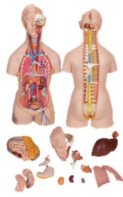
Main Model

VEINS

The right and left brachiocephalic veins are formed posterior to the sternoclavicular (SC) joints by the union of the
internal jugular and subclavian veins. At the level of the inferior border of the 1st right costal cartilage, the brachiocephalic veins unite to form the SVC (Figs. 1.65B and 1.66B).
The left brachiocephalic vein is more than twice as long as the right brachiocephalic vein because it passes from the
left to the right side, anterior to the roots of the three major
branches of the arch of the aorta (Fig. 1.66B). The brachiocephalic veins shunt blood from the head, neck, and upper
limbs to the right atrium.
---------------------------------------------------------------------------------------------------------------------------------------------------------------
Most veins in the anterior cervical region are tributaries of
the IJV, typically the largest vein in the neck (Figs. 8.15 and
8.20). The IJV drains blood from the brain, anterior face,
cervical viscera, and deep muscles of the neck. It commences
at the jugular foramen in the posterior cranial fossa as the
direct continuation of the sigmoid sinus (see Chapter 7).
The IJV ends posterior to the medial end of the clavicle by
uniting with the subclavian vein to form the brachiocephalic
vein. This union is commonly referred to as the venous
angle and is the site where the thoracic duct (left side)
and the right lymphatic trunk (right side) drain lymph collected throughout the body into the venous circulation
(see Fig. 8.48). Throughout its course, the IJV is enclosed by
the carotid sheath (Fig. 8.21).
---------------------------------------------------------------------------------------------------------------------------------------------------------------
The subclavian vein, the continuation of the axillary
vein, begins at the lateral border of the 1st rib and ends when
it unites with the IJV (Fig. 8.24A). The subclavian vein passes
over the 1st rib anterior to the scalene tubercle parallel to the
subclavian artery, but it is separated from it by the anterior
scalene muscle. It usually has only one named tributary, the
EJV (Fig. 8.20).
---------------------------------------------------------------------------------------------------------------------------------------------------------------
The superior vena cava (SVC) returns blood from all
structures superior to the diaphragm, except the lungs and
heart. It passes inferiorly and ends at the level of the 3rd
costal cartilage, where it enters the right atrium of the heart.
The SVC lies in the right side of the superior mediastinum,
anterolateral to the trachea and posterolateral to the ascending aorta. The right phrenic nerve lies between the SVC and
the mediastinal pleura. The terminal half of the SVC is in the
middle mediastinum, where it lies beside the ascending aorta
and forms the posterior boundary of the transverse pericardial sinus (Fig. 1.46).
---------------------------------------------------------------------------------------------------------------------------------------------------------------
The pulmonary trunk, approximately 5 cm long and
3 cm wide, is the arterial continuation of the right ventricle
and divides into right and left pulmonary arteries. The pulmonary trunk and arteries conduct low-oxygen blood to the
lungs for oxygenation (Figs. 1.49A and 1.52B).
---------------------------------------------------------------------------------------------------------------------------------------------------------------
The veins of the posterior abdominal wall are tributaries of
the IVC, except for the left testicular or ovarian vein, which
enters the left renal vein instead of entering the IVC (Fig.
2.99). The IVC, the largest vein in the body, has no valves
except for a variable, non-functional one at its orifi ce in the
right atrium of the heart. The IVC returns poorly oxygenated
blood from the lower limbs, most of the back, the abdominal walls, and the abdominopelvic viscera. Blood from the
abdominal viscera passes through the portal venous system
and the liver before entering the IVC via the hepatic veins.
The inferior vena cava (IVC) begins anterior to the L5
vertebra by the union of the common iliac veins. The union
occurs approximately 2.5 cm to the right of the median plane,
inferior to the aortic bifurcation and posterior to the proximal
part of the right common iliac artery (see Fig. 2.76). The IVC
ascends on the right side of the bodies of the L3–L5 vertebrae and on the right psoas major to the right of the aorta. The IVC leaves the abdomen by passing through the caval
opening in the diaphragm and enters the thorax at the T8
vertebral level. Because it is formed one vertebral level inferior to the aortic bifurcation, and traverses the diaphragm
four vertebral levels superior to the aortic hiatus, the overall
length of the IVC is 7 cm greater than that of the abdominal
aorta, although most of the additional length is intrahepatic.
The IVC collects poorly oxygenated blood from the lower
limbs and non-portal blood from the abdomen and pelvis.
Almost all the blood from the gastrointestinal tract is collected by the hepatic portal system and passes through the
hepatic veins to the IVC.
The tributaries of the IVC correspond to the paired visceral and parietal branches of the abdominal aorta. The
veins that correspond to the unpaired visceral branches of
the aorta are instead tributaries of the hepatic portal vein.
The blood they carry does ultimately enter the IVC via the
hepatic veins, after traversing the liver.
The branches corresponding to the paired visceral
branches of the abdominal aorta include the right suprarenal
vein, the right and left renal veins, and the right gonadal (testicular or ovarian) vein. The left suprarenal and gonadal veins
drain indirectly into the IVC because they are tributaries of
the left renal vein.
Paired parietal branches of the IVC include the inferior
phrenic veins, the 3rd (L3) and 4th (L4) lumbar veins, and
the common iliac veins. The ascending lumbar and azygos
veins connect the IVC and SVC, either directly or indirectly providing collateral pathways (see the blue box “Collateral
Routes for Abdominopelvic Venous Blood” on p. 319).
---------------------------------------------------------------------------------------------------------------------------------------------------------------
Several renal veins drain each kidney and unite in a variable
fashion to form the right and left renal veins; these veins lie
anterior to the right and left renal arteries. The longer left renal
vein receives the left suprarenal vein, the left gonadal (testicular
or ovarian) vein, and a communication with the ascending lumbar
vein; it then traverses the acute angle between the SMA anteriorly
and the aorta posteriorly (see the blue box “Renal Vein Entrapment Syndrome” on p. 298). Each renal vein drains into the IVC.