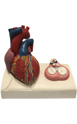
Main Model

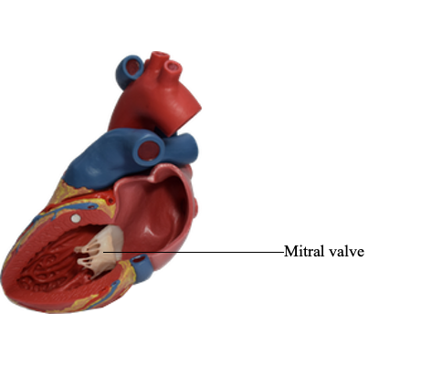
Mitral valve

The mitral valve is located posterior to the sternum at the
level of the 4th costal cartilage. The mitral valve has
two cusps, anterior and posterior. Each of its cusps
receives tendinous cords from more than one papillary muscle.
These muscles and their cords support the mitral valve,
allowing the cusps to resist the pressure developed during
contractions (pumping) of the left ventricle. The cords
become taut just before and during systole, preventing the
cusps from being forced into the left atrium. As it traverses the left ventricle, the bloodstream undergoes two right
angle turns, which together result in a 180° change in
direction. This reversal of flow takes place around the anterior
cusp of the mitral valve.