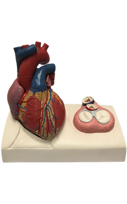
Main Model

Right ventricle

Right Ventricle
The right ventricle forms the largest part of the anterior surface
of the heart, a small part of the diaphragmatic surface and almost the entire
inferior border of the heart. Superiorly, it tapers into an arterial
cone, the conus arteriosus (infundibulum), which leads into the pulmonary trunk. The interior of the right ventricle has irregular muscular
elevations (trabeculae carneae). A thick muscular ridge, the supraventricular
crest, separates the ridged muscular wall of the inflow part of the chamber
from the smooth wall of the conus arteriosus, or outflow part. The inflow part of
the ventricle receives blood from the right atrium through the right atrioventricular (tricuspid) orifice, located posterior to the body of the sternum
at the level of the 4th and 5th intercostal spaces. The
right atrioventricular orifice is surrounded by one of the fibrous rings of the fibrous skeleton of the heart. The fibrous ring keeps the caliber of the orifice constant (large enough
to admit the tips of three fingers), resisting the dilation that might
otherwise result from blood being forced through it at varying pressures.
The tricuspid valve guards the right atrioventricular orifice. The bases of the valve cusps are attached to the fibrous ring around
the orifice. Because the fibrous ring maintains the caliber of the orifice, the
attached valve cusps contact each other in the same way with each heartbeat. Tendinouscords
(Latin chordae tendineae) attach
to the free edges and ventricular surfaces of
the anterior, posterior and septal cusps, much
like the cords attaching to a parachute. The tendinous cords arise from the apices of papillary
muscles, which are conical muscular
projections with bases attached to the
ventricular wall. The papillary muscles begin to contract before contraction of the right ventricle,
tightening the tendinous cords and drawing the
cusps together. Because the cords are attached
to adjacent sides of two cusps, they prevent separation of the cusps and their inversion when tension is applied to the tendinous cords and maintained throughout
ventricular contraction (systole) - that is, the cusps of the
tricuspid valve are prevented from prolapsing
(being driven into the right atrium) as
ventricular pressure rises. Thus, regurgitation of blood
(backward flow of blood) from the right ventricle back into the right atrium is blocked during ventricular systole by the valve cusps.
Three
papillary muscles in the right ventricle correspond to the cusps of the
tricuspid valve:
1.
The anterior papillary muscle, the largest and most prominent of the three,
arises from the anterior wall of the right ventricle; its tendinous cords
attach to the anterior and posterior cusps of the tricuspid valve.
2. The posterior papillary muscle, smaller than the anterior muscle, may consist of several parts; it arises from the inferior wall of the right ventricle, and its tendinous cords attach to the posterior and septal cusps of the tricuspid valve.
3. The septal papillary muscle arises from the interventricular septum, and its tendinous cords attach to the anterior and septal cusps of the tricuspid valve.
The
interventricular septum, composed of muscular and membranous parts, is a
strong, obliquely placed partition between the right and left ventricles, forming part of the walls of each. Because of the much higher
blood pressure in the left ventricle, the muscular part of the interventricular septum, which forms
the majority of the septum, has the thickness of the remainder of the wall of
the left ventricle (two to three times as thick as the wall of the right
ventricle) and bulges into the cavity of the right ventricle. Superiorly and
posteriorly, a thin membrane, part of the fibrous skeleton of the heart, forms the much smaller membranous part of the interventricular septum. On the right side,
the septal cusp of the tricuspid valve is attached to the middle of
this membranous part of the fibrous skeleton. This means that inferior to the cusp,
the membrane is an interventricular septum, but superior to the cusp it is an atrioventricular septum, separating
the right atrium from the left ventricle.
The
septomarginal trabecula (moderator band) is a curved muscular bundle that traverses
the right ventricular chamber from the inferior part of
the interventricular septum to the base of the anterior papillary
muscle. This trabecula is important because it
carries part of the right branch of the atrioventricular bundle, a part of the conducting system of the heart to the anterior
papillary muscle.
This “shortcut” across the chamber seems to
facilitate conduction time, allowing coordinated contraction of the anterior papillary muscle.
The right atrium contracts when the right ventricle is empty and relaxed; thus blood is forced through this orifice into the right ventricle, pushing the cusps of the tricuspid valve aside like curtains. The inflow of blood into the right ventricle (inflow tract) enters posteriorly; and when the ventricle contracts, the outflow of blood into the pulmonary trunk (outflow tract) leaves superiorly and to the left. Consequently, the blood takes a U - shaped path through the right ventricle, changing direction about 140°. This change in direction is accommodated by the supraventricular crest, which deflects the incoming flow into the main cavity of the ventricle, and the outgoing flow into the conus arteriosus toward the pulmonary orifice. The inflow (atrioventricular or tricuspid) orifice and outflow (pulmonary) orifice are approximately 2 cm apart. The pulmonary valve at the apex of the conus arteriosus is at the level of the left 3rd costal cartilage.