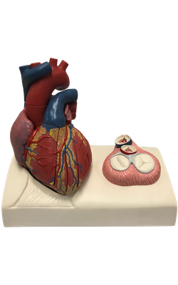
Heart Model

Admin

Main Model


Apex of heart

The apex of the heart is formed by the inferolateral part of the left ventricle. It lies posterior to the left 5th intercostal space in adults, usually approximately 9 cm (a hand’s breadth) from the median plane. It remains motionless throughout the cardiac cycle. It is where the sounds of mitral valve closure are maximal (apex beat); the apex underlies the site where the heartbeat may be auscultated on the thoracic wall.

  • Brachiocephalic trunc
  • Common carotid artery
  • Ascending aorta
  • Aortic arch
  • Pulmonary trunk
  • Subclavian artery
  • Anterior interventricular branch
  • Left coronary artery
  • Great cadiac vein
  • Left pulmonary artery
  • Mitral valve
  • Right pulmonary artery
  • Left atrium
  • Muscular part of interventricular septum
  • Pulmonary valve
  • Tricuspid valve
  • Right atrium
  • Papillary muscles
  • Superior vena cava
  • Left brachiocephalic vein
  • Right brachiocephalic vein
  • Inferior vena cava
  • Right fibrous ring
  • Tricuspid valve (Septal cusp)
  • Tricuspid valve (Posterior cusp)
  • Tricuspid valve (Anterior cusp)
  • Mitral valve (Posterior cusp)
  • Mitral valve (Anterior cusp)
  • Right fibrous trigone
  • Left fibrous ring
  • Left fibrous trigone
  • Aortic valve (Left semilunar cusp)
  • Aortic valve (Posterior semilunar cusp)
  • Aortic valve (Right semilunar cusp)
  • Pulmonary valve (Left semilunar cusp)
  • Pulmonary valve (Anterior semilunar cusp)
  • Pulmonary valve (Right semilunar cusp)
  • Coronary sinus
  • Right coronary artery
  • Posterior interventricular branch
  • Right ventricle
  • Left ventricle
  • Apex of heart
  • Middle cardiac vein

Copyright 2018, Chulabhorn International College of Medicine

www.cicm.tu.ac.th