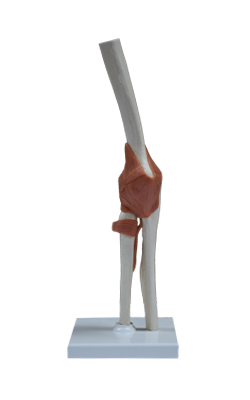
Main Model
Posterior : Radius
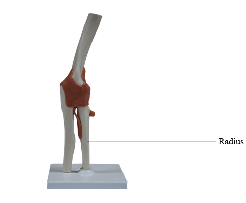
Radius
The radius is the lateral and shorter of the two forearm bones. Its proximal end includes a short head, neck, and medially directed tuberosity. Proximally, the smooth superior aspect of the discoid head of the radius is concave for articulation with the capitulum of the humerus during flexion and extension of the elbow joint. The head also articulates peripherally with the radial notch of the ulna; thus the head is covered with articular cartilage.
The neck of the radius is a constriction distal to the head. The oval radial tuberosity is distal to the medial part of the neck, and demarcates the proximal end (head and neck) of the radius from the shaft.
The shaft of the radius, in contrast to that of the ulna, gradually enlarges as it passes distally. The distal end of the radius is essentially four sided when sectioned transversely. Its medial aspect forms a concavity, the ulnar notch, which accommodates the head of the ulna. Its lateral aspect becomes increasingly ridge-like, terminating distally in the radial styloid process.
Projecting dorsally, the dorsal tubercle of the radius lies between otherwise shallow grooves for the passage of the tendons of forearm muscles. The radial styloid process is larger than the ulnar styloid process, and extends farther distally. This relationship is of clinical importance when the ulna and/or the radius is fractured.
Most of the length of the shafts of the radius and ulna are essentially triangular in cross section, with a rounded, superficially directed base and an acute, deeply directed apex. The apex is formed by a section of the sharp interosseous border of the radius or ulna that connects to the thin, fibrous interosseous membrane of the forearm. The majority of the fibers of the interosseous membrane run an oblique course, passing inferiorly from the radius as they extend medially to the ulna. Thus, they are positioned to transmit forces received by the radius (via the hands) to the ulna for transmission to the humerus.