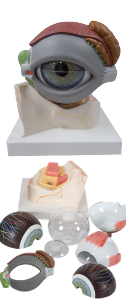
Main Model
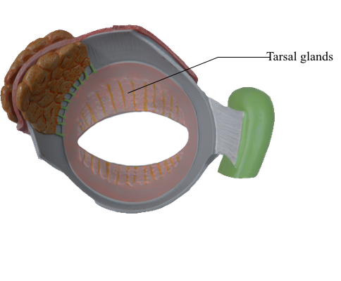
EYELID : 8 Tarsal glands
Eyelids
The eyelids are moveable folds that cover the eyeball anteriorly when closed, thereby protecting it from injury and excessive light. They also keep the cornea moist by spreading the lacrimal fluid. The eyelids are covered externally by thin skin and internally by transparent mucous membrane, the palpebral conjunctiva. This part of the conjunctiva is reflected onto the eyeball, where it is continuous with the bulbar conjunctiva. This part of the conjunctiva is thin and transparent and attaches loosely to the anterior surface of the eyeball. The bulbar conjunctiva, loose and wrinkled over the sclera (where it contains small, visible blood vessels), is adherent to the periphery of the cornea. The lines of reflection of the palpebral conjunctiva onto the eyeball form deep recesses, the superior and inferior conjunctival fornices.
The conjunctival sac is the space bound by the palpebral and bulbar conjunctivae; it is a closed space when the eyelids are closed, but opens via an anterior aperture, the palpebral fissure (Latin rima palpebrae, the gap between the eyelids), when the eye is open (eyelids are parted). The conjunctival sac is a specialized form of mucosal "bursa" that enables the eyelids to move freely over the surface of the eyeball as they open and close.
The superior (upper) and inferior (lower) eyelids are strengthened by dense bands of connective tissue, the superior and inferior tarsi (singular = tarsus), which form the "skeleton" of the eyelids. Fibers of the palpebral portion of the orbicularis oculi (the sphincter of the palpebral fissure) are in the connective tissue superficial to the tarsi and deep to the skin of the eyelids. Embedded in the tarsi are tarsal glands that produce a lipid secretion that lubricates the edges of the eyelids and prevents them from sticking together when they close. The lipid secretion also forms a barrier that lacrimal fluid does not cross when produced in normal amounts. When production is excessive, it spills over the barrier onto the cheeks as tears.
The eyelashes (Latin cilia) are in the margins of the eyelids. The large sebaceous glands associated with the eyelashes are ciliary glands. The junctions of the superior and inferior eyelids make up the medial and lateral palpebral commissures, defining the medial and lateral angles of the eye (Greek kanthos, corner of eye), or canthi.
Between the nose and the medial angle of the eye is the medial palpebral ligament, which connects the tarsi to the medial margin of the orbit. The orbicularis oculi originates and inserts onto this ligament. A similar lateral palpebral ligament attaches the tarsi to the lateral margin of the orbit, but it does not provide for direct muscle attachment.
The orbital septum is a fibrous membrane that spans from the tarsi to the margins of the orbit, where it becomes continuous with the periosteum. It keeps the orbital fat contained and, owing to its continuity with the periorbita, can limit the spread of infection to and from the orbit. The septum constitutes in large part the posterior fascia of the orbicularis oculi muscle.