Main Model
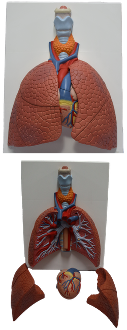
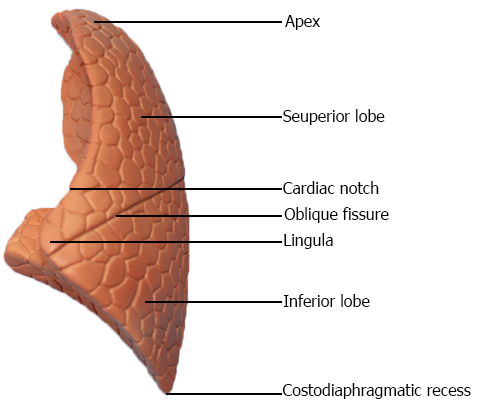
LUNGS : Left lung
Lungs
The lungs are the vital organs of respiration. Their main function is to oxygenate the blood by bringing inspired air into close relation with the venous blood in the pulmonary capillaries. Although cadaveric lungs may be shrunken, firm or hard, and discolored, healthy lungs in living people are normally light, soft, and spongy, and fully occupy the pulmonary cavities. They are also elastic and recoil to approximately one third their size when the thoracic cavity is opened. The lungs are separated from each other by the mediastinum. Each lung has:
• An apex, the blunt superior end of the lung ascending above the level of the 1st rib into the root of the neck; the apex is covered by cervical pleura.
• A base, the concave inferior surface of the lung, opposite the apex, resting on and accommodating the ipsilateral dome of the diaphragm.
• Two or three lobes, created by one or two fissures.
• Three surfaces (costal, mediastinal, and diaphragmatic).
• Three borders (anterior, inferior, and posterior).
The right lung features right oblique and horizontal fissures that divide it into three right lobes: superior, middle, and inferior. The right lung is larger and heavier than the left, but it is shorter and wider because the right dome of the diaphragm is higher and the heart and pericardium bulge more to the left. The anterior border of the right lung is relatively straight. The left lung has a single left oblique fissure dividing it into two left lobes, superior and inferior. The anterior border of the left lung has a deep cardiac notch, an indentation consequent to the deviation of the apex of the heart to the left side. This notch primarily indents the antero-inferior aspect of the superior lobe. This indentation often shapes the most inferior and anterior part of the superior lobe into a thin, tongue-like process, the lingula (Latin dim. of lingua, tongue), which extends below the cardiac notch and slides in and out of the costomediastinal recess during inspiration and expiration.
The lungs of an embalmed cadaver, usually firm to the touch, demonstrate impressions formed by structures adjacent to them, such as the ribs, heart, and great vessels. These markings provide clues to the relationships of the lungs; however, only the cardiac impressions are evident during surgery or in fresh cadaveric or postmortem specimens.
The costal surface of the lung is large, smooth, and convex. It is related to the costal pleura, which separates it from the ribs, costal cartilages, and innermost intercostal muscles. The posterior part of the costal surface is related to the bodies of the thoracic vertebrae and is sometimes referred to as the vertebral part of the costal surface.
The mediastinal surface of the lung is concave because it is related to the middle mediastinum, which contains the pericardium and heart. The mediastinal surface includes the hilum, which receives the root of the lung. If embalmed, there is a groove for the esophagus and a cardiac impression for the heart on the mediastinal surface of the right lung. Because two thirds of the heart lies to the left of the midline, the cardiac impression on the mediastinal surface of the left lung is much larger. This surface of the left lung also features a prominent, continuous groove for the arch of the aorta and the descending aorta as well as a smaller area for the esophagus.
The diaphragmatic surface of the lung, which is also concave, forms the base of the lung, which rests on the dome of the diaphragm. The concavity is deeper in the right lung because of the higher position of the right dome, which overlies the liver. Laterally and posteriorly, the diaphragmatic surface is bounded by a thin, sharp margin (inferior border) that projects into the costodiaphragmatic recess of the pleura.
The anterior border of the lung is where the costal and mediastinal surfaces meet anteriorly and overlap the heart. The cardiac notch indents this border of the left lung. The inferior border of the lung circumscribes the diaphragmatic surface of the lung and separates this surface from the costal and mediastinal surfaces. The posterior border of the lung is where the costal and mediastinal surfaces meet posteriorly; it is broad and rounded and lies in the cavity at the side of the thoracic region of the vertebral column.
The lungs are attached to the mediastinum by the roots of the lungs - that is, the bronchi (and associated bronchial vessels), pulmonary arteries, superior and inferior pulmonary veins, the pulmonary plexuses of nerves (sympathetic, parasympathetic, and visceral afferent fibers), and lymphatic vessels. If the lung root is sectioned before the (medial to) branching of the main (primary) bronchus and pulmonary artery, its general arrangement is
• Pulmonary artery: superiormost on left (the superior lobar or "eparterial" bronchus may be superiormost on the right).
• Superior and inferior pulmonary veins: anteriormost and inferiormost, respectively.
• Main bronchus: against and approximately in the middle of the posterior boundary, with the bronchial vessels coursing on its outer surface (usually on posterior aspect at this point).
The hilum of the lung is a wedge-shaped area on the mediastinal surface of each lung through which the structures forming the root of the lung pass to enter or exit the lung. The hilum ("doorway") can be likened to the area of earth where a plant's roots enter the ground. Medial to the hilum, the lung root is enclosed within the area of continuity between the parietal and the visceral layers of pleura - the pleural sleeve (mesopneumonium).
Inferior to the root of the lung, this continuity between parietal and visceral pleura forms the pulmonary ligament, extending between the lung and the mediastinum, immediately anterior to the esophagus. The pulmonary ligament consists of a double layer of pleura separated by a small amount of connective tissue. When the root of the lung is severed and the lung is removed, the pulmonary ligament appears to hang from the root. To visualize the root of the lung, the pleural sleeve surrounding it, and the pulmonary ligament hanging from it, put on an extra-large lab coat and abduct your upper limb. Your forearm is comparable to the root of the lung, and the coat sleeve represents the pleural sleeve surrounding it. The pulmonary ligament is comparable to the slack of the sleeve as it hangs from your forearm; and your wrist, hand, and abducted fingers represent the branching structures of the root - the bronchi and pulmonary vessels.