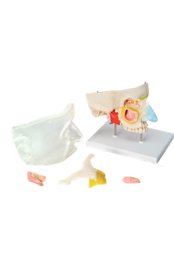
Main Model

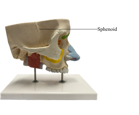
Anterior : Sphenoid

External Surface of Cranial Base
The cranial base (basicranium) is the inferior portion of the neurocranium (floor of the cranial cavity) and viscerocranium minus the mandible. The external surface of the cranial base features the alveolar arch of the maxillae (the free border of the alveolar processes surrounding and supporting the maxillary teeth); the palatine processes of the maxillae; and the palatine, sphenoid, vomer, temporal, and occipital bones.
The hard palate (bony palate) is formed by the palatal processes of the maxillae anteriorly and the horizontal plates of the palatine bones posteriorly. The free posterior border of the hard palate projects posteriorly in the median plane as the posterior nasal spine. Posterior to the central incisor teeth is the incisive foramen, a depression in the midline of the bony palate into which the incisive canals open.
The right and left nasopalatine nerves pass from the nose through a variable number of incisive canals and foramina (they may be bilateral or merged into a single formation). Posterolaterally are the greater and lesser palatine foramina. Superior to the posterior edge of the palate are two large openings: the choanae (posterior nasal apertures), which are separated from each other by the vomer (Latin plowshare), a flat unpaired bone of trapezoidal shape that forms a major part of the bony nasal septum.
Wedged between the frontal, temporal, and occipital bones is the sphenoid, an irregular unpaired bone that consists of a body and three pairs of processes: greater wings, lesser wings, and pterygoid processes. The greater and lesser wings of the sphenoid spread laterally from the lateral aspects of the body of the bone. The greater wings have orbital, temporal, and infratemporal surfaces apparent in facial, lateral, and inferior views of the exterior of the cranium and cerebral surfaces seen in internal views of the cranial base. The pterygoid processes, consisting of lateral and medial pterygoid plates, extend inferiorly on each side of the sphenoid from the junction of the body and greater wings.
The groove for the cartilaginous part of the pharyngotympanic (auditory) tube lies medial to the spine of the sphenoid, inferior to the junction of the greater wing of the sphenoid and the petrous (Latin rock-like) part of the temporal bone. Depressions in the squamous (Latin flat) part of the temporal bone, called the mandibular fossae, accommodate the mandibular condyles when the mouth is closed. The cranial base is formed posteriorly by the occipital bone, which articulates with the sphenoid anteriorly.
The four parts of the occipital bone are arranged around the foramen magnum, the most conspicuous feature of the cranial base. The major structures passing through this large foramen are: the spinal cord (where it becomes continuous with the medulla oblongata of the brain); the meninges (coverings) of the brain and spinal cord: the vertebral arteries; the anterior and posterior spinal arteries; and the spinal accessory nerve (CN XI). On the lateral parts of the occipital bone are two large protuberances, the occipital condyles, by which the cranium articulates with the vertebral column.
The large opening between the occipital bone and the petrous part of the temporal bone is the jugular foramen, from which the internal jugular vein (IJV) and several cranial nerves (CN IX - CN XI) emerge from the cranium. The entrance to the carotid canal for the internal carotid artery is just anterior to the jugular foramen. The mastoid processes provide for muscle attachments. The stylomastoid foramen, transmitting the facial nerve (CN VII) and stylomastoid artery, lies posterior to the base of the styloid process.
Internal Surface of Cranial Base
The internal surface of the cranial base (Latin basis cranii interna) has three large depressions that lie at different levels: the anterior, middle, and posterior cranial fossae, which form the bowl-shaped floor of the cranial cavity. The anterior cranial fossa is at the highest level, and the posterior cranial fossa is at the lowest level.