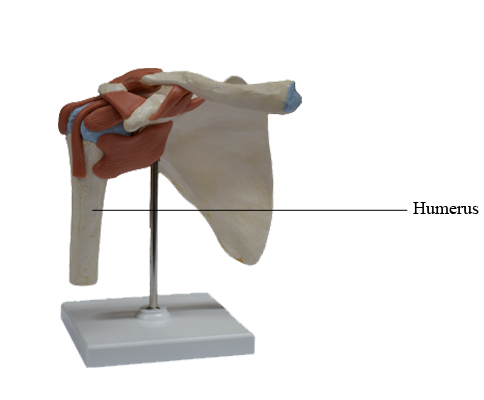
Main Model

Anterior : Humerus

Humerus
The humerus (arm bone), the largest bone in the upper limb, articulates with the scapula at the glenohumeral joint, and the radius and ulna at the elbow joint. The proximal end of the humerus has a head, surgical and anatomical necks, and greater and lesser tubercles. The spherical head of the humerus articulates with the glenoid cavity of the scapula. The anatomical neck of the humerus is formed by the groove circumscribing the head and separating it from the greater and lesser tubercles. It indicates the line of attachment of the glenohumeral joint capsule. The surgical neck of the humerus, a common site of fracture, is the narrow part distal to the head and tubercles.
The junction of the head and neck with the shaft of the humerus is indicated by the greater and lesser tubercles, which provide attachment and leverage to some scapulohumeral muscles. The greater tubercle is at the lateral margin of the humerus, whereas the lesser tubercle projects anteriorly from the bone. The intertubercular sulcus (bicipital groove) separates the tubercles, and provides protected passage for the slender tendon of the long head of the biceps muscle.
The shaft of the humerus has two prominent features: the deltoid tuberosity laterally, for attachment of the deltoid muscle, and the oblique radial groove (groove for radial nerve, spiral groove) posteriorly, in which the radial nerve and profunda brachii artery lie as they pass anterior to the long head and between the medial and the lateral heads of the triceps brachii muscle. The inferior end of the humeral shaft widens as the sharp medial and lateral supra-epicondylar (supracondylar) ridges form, and then end distally in the especially prominent medial epicondyle and the lateral epicondyle, providing for muscle attachment.
The distal end of the humerus - including the trochlea, capitulum, olecranon, coronoid, and radial fossae - makes up the condyle of the humerus. The condyle has two articular surfaces: a lateral capitulum (Latin little head) for articulation with the head of the radius, and a medial, spool-shaped or pulley-like trochlea (Latin pulley) for articulation with the proximal end (trochlear notch) of the ulna. Two hollows, or fossae, occur back to back superior to the trochlea, making the condyle quite thin between the epicondyles. Anteriorly, the coronoid fossa receives the coronoid process of the ulna during full flexion of the elbow. Posteriorly, the olecranon fossa accommodates the olecranon of the ulna during full extension of the elbow. Superior to the capitulum anteriorly, a shallower radial fossa accommodates the edge of the head of the radius when the forearm is fully flexed.