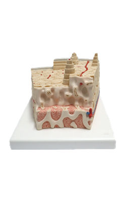
Main Model

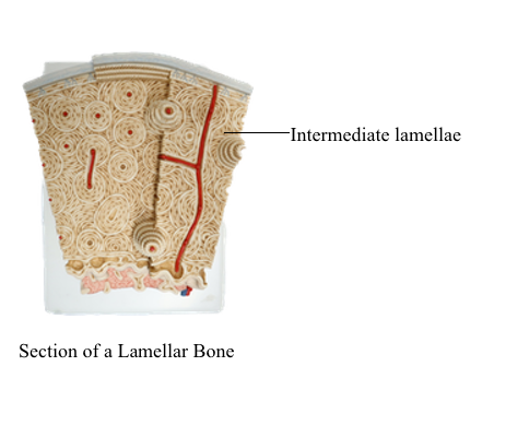
Intermediate lamellae

Microscopic Structure of Mature Bone
Two types of bone are identified on the basis of the microscopic three-dimensional arrangement of the collagen fibers:
1. Lamellar or compact bone, typical of the mature bone, displays a regular alignment of collagen fibers. This bone is mechanically strong and forms slowly.
2. Woven bone, observed in the developing bone, is characterized by an irregular alignment of collagen fibers. This bone is mechanically weak, is formed rapidly and is then replaced by lamellar bone. Woven bone is produced during the repair of a bone fracture.
The lamellar bone consists of lamellae, largely composed of bone matrix, a mineralized substance deposited in layers or lamellae, and osteocytes, each one occupying a cavity or lacuna with radiating and branching canaliculi that penetrate the lamellae of adjacent lacunae.
The lamellar bone displays four distinct patterns:
1. The osteons or haversian systems, formed by concentrically arranged lamellae around a longitudinal vascular channel. About 4 to 20 lamellae are concentrically arranged around the haversian canal.
2. The interstitial lamellae, observed between osteons and separated from them by a thin layer known as the cement line.
3. The outer circumferential lamellae, visualized at the external surface of the compact bone under the periosteum.
4. The inner circumferential lamellae, seen on the internal surface subjacent to the endosteum.
The vascular channels in compact bone have two orientations with respect to the lamellar structures:
1. The longitudinal haversian canal, housing capillaries and postcapillary venules in the center of the osteon.
Blood vessels in a haversian canal run in a direction parallel to the bone shaft.
2. The transverse or oblique Volkmann's canals, connecting haversian canals with one another, containing blood vessels derived from the bone marrow and some from the periosteum.
Blood vessels in Volkmann's canal run in a direction perpendicular/oblique to the haversian canal.