Main Model
Anterior : Maxillary sinus

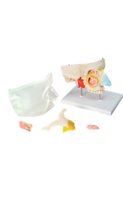
Paranasal Sinuses
The paranasal sinuses are air-filled extensions of the respiratory part of the nasal cavity into the following cranial bones: frontal, ethmoid, sphenoid, and maxilla. They are named according to the bones in which they are located. The sinuses continue to invade the surrounding bone, and marked extensions are common in the crania of older individuals.
Maxillary Sinuses
The maxillary sinuses are the largest of the paranasal sinuses. They occupy the bodies of the maxillae and communicate with the middle nasal meatus.
• The apex of the maxillary sinus extends toward and often into the zygomatic bone.
• The base of the maxillary sinus forms the inferior part of the lateral wall of the nasal cavity.
• The roof of the maxillary sinus is formed by the floor of the orbit.
• The floor of the maxillary sinus is formed by the alveolar part of the maxilla. The roots of the maxillary teeth, particularly the first two molars, often produce conical elevations in the floor of the sinus.
Each maxillary sinus drains by one or more openings, the maxillary ostium (ostia), into the middle nasal meatus of the nasal cavity by way of the semilunar hiatus.
The arterial supply of the maxillary sinus is mainly from superior alveolar branches of the maxillary artery; however, branches of the descending and greater palatine arteries supply the floor of the sinus. Innervation of the maxillary sinus is from the anterior, middle, and posterior superior alveolar nerves, which are branches of the maxillary nerve.