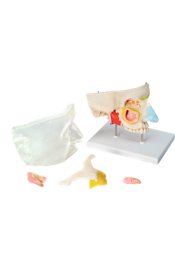
Main Model
Posterior : Anterior and middle ethmoidal cells
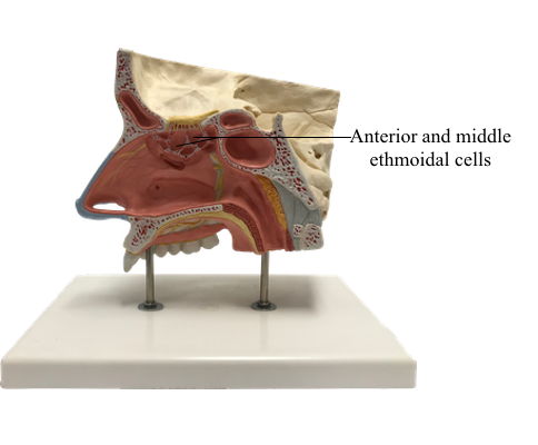
Paranasal Sinuses
The paranasal sinuses are air-filled extensions of the respiratory part of the nasal cavity into the following cranial bones: frontal, ethmoid, sphenoid, and maxilla. They are named according to the bones in which they are located. The sinuses continue to invade the surrounding bone, and marked extensions are common in the crania of older individuals.
Ethmoidal Cells
The ethmoidal cells (sinuses) are small invaginations of the mucous membrane of the middle and superior nasal meatus into the ethmoid bone between the nasal cavity and the orbit. The ethmoidal cells usually are not visible in plain radiographs before 2 years of age but are recognizable in CT scans. The anterior ethmoidal cells drain directly or indirectly into the middle nasal meatus through the ethmoidal infundibulum. The middle ethmoidal cells open directly into the middle meatus and are sometimes called “bullar cells” because they form the ethmoidal bulla, a swelling on the superior border of the semilunar hiatus. The posterior ethmoidal cells open directly into the superior meatus. The ethmoidal cells are supplied by the anterior and posterior ethmoidal branches of the nasociliary nerves (CN V1).