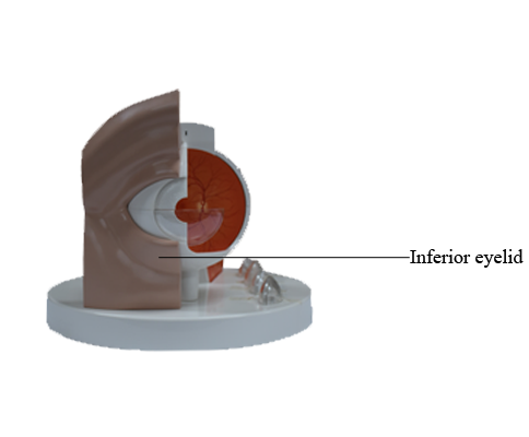
Main Model
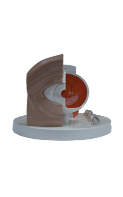
Anterior : Inferior eyelid
Eyelids and Lacrimal Apparatus
The eyelids and lacrimal fluid, secreted by the lacrimal
glands, protect the cornea and eyeballs from injury and irritation (e.g., by dust and small particles).
Eyelids
The eyelids are moveable folds that cover the eyeball anteriorly when closed, thereby protecting it from injury and excessive light. They also keep the cornea moist by spreading the lacrimal fluid. The eyelids are covered externally by thin skin
and internally by transparent mucous membrane, the palpebral conjunctiva. This part of the conjunctiva is reflected onto the eyeball, where it is continuous with
the bulbar conjunctiva. This part of the conjunctiva is thin and transparent and attaches loosely to the anterior surface
of the eyeball. The bulbar conjunctiva, loose and wrinkled
over the sclera (where it contains small, visible blood vessels), is adherent to the periphery of the cornea.
The lines of reflection of the palpebral conjunctiva onto the
eyeball form deep recesses, the superior and inferior conjunctival fornices.
The conjunctival sac is the space bound by the palpebral
and bulbar conjunctivae; it is a closed space when the eyelids
are closed, but opens via an anterior aperture, the palpebral
fissure (Latin rima palpebrae, the gap between the eyelids),
when the eye is open (eyelids are parted). The
conjunctival sac is a specialized form of mucosal "bursa" that
enables the eyelids to move freely over the surface of the
eyeball as they open and close.
The superior (upper) and inferior (lower) eyelids are
strengthened by dense bands of connective tissue, the superior and inferior tarsi (singular = tarsus), which form the "skeleton" of the eyelids. Fibers of
the palpebral portion of the orbicularis oculi (the sphincter of the palpebral fissure) are in the connective tissue
superficial to the tarsi and deep to the skin of the eyelids. Embedded in the tarsi are tarsal glands that produce a lipid secretion that lubricates the edges of the eyelids and
prevents them from sticking together when they close. The lipid secretion also forms a barrier that lacrimal fluid does not cross
when produced in normal amounts. When production is excessive, it spills over the barrier onto the cheeks as tears.
The eyelashes (Latin cilia) are in the margins of the eyelids.
The large sebaceous glands associated with the eyelashes are
ciliary glands. The junctions of the superior and inferior eyelids make up the medial and lateral palpebral commissures, defining the medial and lateral angles of the eye
(Greek kanthos, corner of eye), or canthi.
Between the nose and the medial angle of the eye is the
medial palpebral ligament, which connects the tarsi to the
medial margin of the orbit. The orbicularis oculi
originates and inserts onto this ligament. A similar lateral
palpebral ligament attaches the tarsi to the lateral margin of
the orbit, but it does not provide for direct muscle attachment.
The orbital septum is a fibrous membrane that spans
from the tarsi to the margins of the orbit, where it becomes
continuous with the periosteum. It
keeps the orbital fat contained and, owing to its continuity
with the periorbita, can limit the spread of infection to and
from the orbit. The septum constitutes in large part the posterior fascia of the orbicularis oculi muscle.
Lacrimal Apparatus
The lacrimal apparatus consists of the:
• Lacrimal gland: secretes lacrimal fluid, a watery physiological saline containing the bacteriocidal enzyme lysozyme. The fluid moistens and lubricates the surfaces of
the conjunctiva and cornea and provides some nutrients and dissolved oxygen to the cornea; when produced in
excess, the overflowing fluid constitutes tears.
• Excretory ducts of lacrimal gland: convey lacrimal
fluid from the lacrimal glands to the conjunctival sac.
• Lacrimal canaliculi (Latin small canals): commence at a
lacrimal punctum (opening) on the lacrimal papilla
near the medial angle of the eye and drain lacrimal fluid
from the lacrimal lake (Latin lacus lacrimalis; a triangular space at the medial angle of the eye where the tears collect) to the lacrimal sac (dilated superior part of the
nasolacrimal duct).
• Nasolacrimal duct: conveys the lacrimal fluid to the inferior nasal meatus (part of the nasal cavity inferior to the
inferior nasal concha.
The lacrimal gland, almond shaped and approximately 2 cm
long, lies in the fossa for the lacrimal gland in the superolateral part of each orbit. The
gland is divided into a superior orbital and inferior palpebral parts by the lateral expansion of the tendon of the levator palpebrae superioris. Accessory lacrimal
glands may also be present, sometimes in the middle part
of the eyelid, or along the superior or inferior fornices of the
conjunctival sac. They are more numerous in the superior
eyelid than in the inferior eyelid.
Production of lacrimal fluid is stimulated by parasympathetic impulses from CN VII. It is secreted through 8-12
excretory ducts, which open into the lateral part of the superior conjunctival fornix of the conjunctival sac. The fluid
flows inferiorly within the sac under the influence of gravity.
When the cornea becomes dry, the eye blinks. The eyelids
come together in a lateral to medial sequence pushing a film
of fluid medially over the cornea, somewhat like windshield wipers. In this way, lacrimal fluid, containing foreign material such as dust is pushed toward the medial angle of the
eye, accumulating in the lacrimal lake from which it drains
by capillary action through the lacrimal puncta and lacrimal
canaliculi to the lacrimal sac.
From this sac, the fluid passes to the inferior nasal meatus
of the nasal cavity through the nasolacrimal duct. It drains
posteriorly across the floor of the nasal cavity to the nasopharynx and is eventually swallowed. In addition to cleansing
particles and irritants from the conjunctival sac, lacrimal fluid
provides the cornea with nutrients and oxygen.
The nerve supply of the lacrimal gland is both sympathetic and parasympathetic. The presynaptic
parasympathetic secretomotor fibers are conveyed from the
facial nerve by the greater petrosal nerve and then by the
nerve of the pterygoid canal to the pterygopalatine ganglion,
where they synapse with the cell body of the postsynaptic
fiber. Vasoconstrictive, postsynaptic sympathetic fibers,
brought from the superior cervical ganglion by the internal carotid plexus and deep petrosal nerve, join the parasympathetic fibers to form the nerve of the pterygoid canal and
traverse the pterygopalatine ganglion. The zygomatic nerve
(from the maxillary nerve) brings both types of fibers to the
lacrimal branch of the ophthalmic nerve, by which they enter
the gland.