Main Model
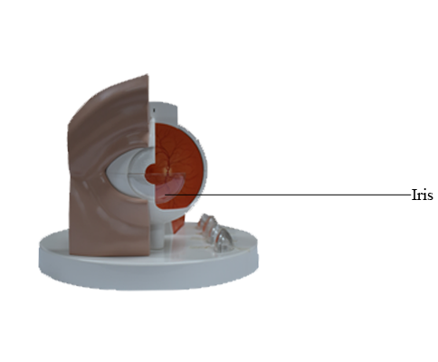
Anterior : Iris
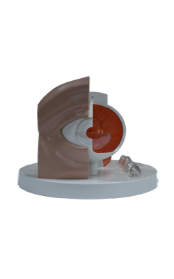
Eyeball
The eyeball contains the optical apparatus of the visual system. It occupies most of the anterior portion of the orbit, suspended by six extrinsic muscles that control its movement, and a fascial suspensory apparatus. It measures approximately 25 mm in diameter. All anatomical structures within the eyeball have a circular or spherical arrangement. The eyeball proper has three layers; however, there is an additional connective tissue layer that surrounds the eyeball, supporting it within the orbit. The connective tissue layer is composed posteriorly of the fascial sheath of the eyeball (bulbar fascia or Tenon capsule), which forms the actual socket for the eyeball, and anteriorly of bulbar conjunctiva. The fascial sheath is the most substantial portion of the suspensory apparatus. A very loose connective tissue layer, the episcleral space (a potential space) lies between the fascial sheath and the outer layer of the eyeball, facilitating movements of the eyeball within the fascial sheath.
The three layers of the eyeball are the:
1. Fibrous layer (outer coat), consisting of the sclera and cornea.
2. Vascular layer (middle coat), consisting of the choroid, ciliary body, and iris.
3. Inner layer (inner coat), consisting of the retina, which has both optic and non-visual parts.
Fibrous Layer of Eyeball
The fibrous layer of the eyeball is the external fibrous skeleton of the eyeball, providing shape and resistance. The sclera is the tough opaque part of the fibrous layer (coat) of the eyeball, covering the posterior five sixths of the eyeball. It provides attachment for both the extrinsic (extra-ocular) and intrinsic muscles of the eye. The anterior part of the sclera is visible through the transparent bulbar conjunctiva as "the white of the eye". The cornea is the transparent part of the fibrous layer covering the anterior one sixth of the eyeball. The convexity of the cornea is greater than that of the sclera, and so it appears to protrude from the eyeball when viewed laterally.
The two parts of the fibrous layer differ primarily in terms of the regularity of arrangement of the collagen fibers of which they are composed and the degree of hydration of each. While the sclera is relatively avascular, the cornea is completely avascular, receiving its nourishment from capillary beds around its periphery and fluids on its external and internal surfaces (lacrimal fluid and aqueous humor, respectively). Lacrimal fluid also provides oxygen absorbed from the air.
The cornea is highly sensitive to touch, its innervation is provided by the ophthalmic nerve (CN V1). Even very small foreign bodies (e.g., dust particles) elicit blinking, flow of tears, and sometimes severe pain. Its nourishment is derived from the capillary beds at its periphery, the aqueous humor, and lacrimal fluid. The latter also provides oxygen absorbed from air. Drying of the corneal surface may cause ulceration.
The corneal limbus is the angle formed by the intersecting curvatures of sclera and cornea at the corneoscleral junction. The junction is a 1-mm-wide, gray, translucent circle that includes numerous capillary loops involved in nourishing the avascular cornea.
Vascular Layer of Eyeball
The middle vascular layer of the eyeball (also called the uvea or uveal tract) consists of the choroid, ciliary body, and iris. The choroid, a dark reddish brown layer between the sclera and retina, forms the largest part of the vascular layer of the eyeball and lines most of the sclera. Within this pigmented and dense vascular bed, larger vessels are located externally (near the sclera). The finest vessels (the capillary lamina of the choroid, or choriocapillaris, an extensive capillary bed) are innermost, adjacent to the avascular light-sensitive layer of the retina, which it supplies with oxygen and nutrients. Engorged with blood in life (it has the highest perfusion rate per gram of tissue of all vascular beds of the body), this layer is responsible for the "red eye" reflection that occurs in flash photography. The choroid attaches firmly to the pigment layer of the retina, but can easily be stripped from the sclera. The choroid is continuous anteriorly with the ciliary body.
The ciliary body, is a ring-like thickening of the layer posterior to the corneoscleral junction, which is muscular as well as vascular. It connects the choroid with the circumference of the iris. The ciliary body provides attachment for the lens. The contraction and relaxation of the circularly arranged smooth muscle of the ciliary body controls the thickness, and therefore the focus, of the lens. Folds on the internal surface of the ciliary body, the ciliary processes, secrete aqueous humor. Aqueous humor fills the anterior segment of the eyeball, the interior of the eyeball anterior to the lens, suspensory ligament, and ciliary body.
The iris, which literally lies on the anterior surface of the lens, is a thin contractile diaphragm with a central aperture, the pupil, for transmitting light. When a person is awake, the size of the pupil varies continually to regulate the amount of light entering the eye. Two involuntary muscles control the size of the pupil: the parasympathetically stimulated, circularly arranged sphincter pupillae decreases its diameter (constrict or contracts the pupil, pupillary miosis), and the sympathetically stimulated, radially arranged dilator pupillae increases its diameter (dilates the pupil). The nature of the pupillary responses is paradoxical: sympathetic responses usually occur immediately, yet it may take up to 20 min for the pupil to dilate in response to low lighting, as in a darkened theater. Parasympathetic responses are typically slower than sympathetic responses, yet parasympathetically stimulated papillary constriction is normally instantaneous. Abnormal sustained pupillary dilation (mydriasis) may occur in certain diseases or as a result of trauma or the use of certain drugs.
Inner Layer of Eyeball
The inner layer of the eyeball is the retina. It is the sensory neural layer of the eyeball. Grossly, the retina consists of two functional parts with distinct locations: an optic part and a non-visual retina. The optic part of the retina is sensitive to visual light rays and has two layers: a neural layer and pigmented layer. The neural layer is light receptive. The pigmented layer consists of a single layer of cells that reinforces the light-absorbing property of the choroid in reducing the scattering of light in the eyeball. The non-visual retina is an anterior continuation of the pigmented layer and a layer of supporting cells. The non-visual retina extends over the ciliary body (ciliary part of retina) and the posterior surface of the iris (iridial part of retina), to the pupillary margin.
Clinically, the internal aspect of the posterior part of the eyeball, where light entering the eyeball is focused, is referred to as the fundus of the eyeball (ocular fundus). The retina of the fundus includes a distinctive circular area called the optic disc (optic papilla), where the sensory fibers and vessels conveyed by the optic nerve (CN II) enter the eyeball. Because it contains no photoreceptors, the optic disc is insensitive to light. Consequently, this part of the retina is commonly called the blind spot.
Just lateral to the optic disc is the macula of the retina or macula lutea (Latin yellow spot). The yellow color of the macula is apparent only when the retina is examined with red-free light. The macula is a small oval area of the retina with special photoreceptor cones that is specialized for acuity of vision. It is not normally observed with an ophthalmoscope (a device for viewing the interior of the eyeball through the pupil). At the center of the macula, a depression, the fovea centralis (Latin central pit), is the area of most acute vision. The fovea is approximately 1.5 mm in diameter; its center, the foveola, does not have the capillary network visible elsewhere deep to the retina.
The optic part of the retina terminates anteriorly along the ora serrata (Latin serrated edge), the irregular posterior border of the ciliary body. Except for the cones and rods of the neural layer, the retina is supplied by the central artery of the retina, a branch of the ophthalmic artery. The cones and rods of the outer neural layer receive nutrients from the capillary lamina of the choroid, or choriocapillaris. It has the finest vessels of the inner surface of the choroid, against which the retina is pressed. A corresponding system of retinal veins unites to form the central vein of the retina.