Main Model
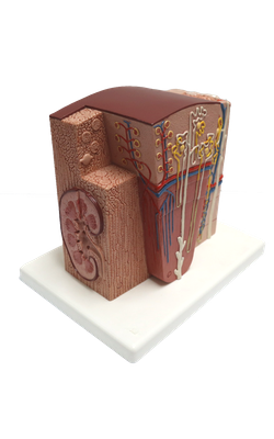
Zone A : Ureter
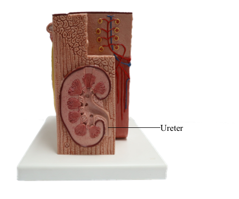
Ureter
The ureters are muscular ducts (25-30 cm long) with narrow lumina that carry urine from the kidneys to the urinary bladder. They run inferiorly from the apices of the renal pelves at the hila of the kidneys, passing over the pelvic brim at the bifurcation of the common iliac arteries. They then run along the lateral wall of the pelvis and enter the urinary bladder.
The ureters are muscular ducts (25-30 cm long) with narrow lumina that carry urine from the kidneys to the urinary bladder. They run inferiorly from the apices of the renal pelves at the hila of the kidneys, passing over the pelvic brim at the bifurcation of the common iliac arteries. They then run along the lateral wall of the pelvis and enter the urinary bladder.
The abdominal parts of the ureters adhere closely to the parietal peritoneum and are retroperitoneal throughout their course. From the back, the surface marking of the ureter is a line joining a point 5 cm lateral to the L1 spinous process and the posterior superior iliac spine. The ureters occupy a sagittal plane that intersects the tips of the transverse processes of the lumbar vertebrae. When examining the ureters radiographically using contrast medium, the ureters normally demonstrate relative constrictions in three places:
1. At the junction of the ureters and renal pelves
2. Where the ureters cross the brim of the pelvic inlet and
3. During their passage through the wall of the urinary bladder.
These constricted areas are potential sites of obstruction by ureteric stones (calculi). Congenital anomalies of the ureters are fairly common.
Ureter
The ureters are muscular tubes, 25-30 cm long and 5-10 mm in diameter, leading from the renal pelvis to the urinary bladder. The wall of the ureter consists of three layers: mucosa, muscularis, and adventitia of connective tissue.
The mucosa is composed of the transitional epithelium and lamina propria. The transitional epithelium, which rests upon a basement membrane, consists of several layers of epithelial cells. The underlying lamina propria is a loose connective tissue containing many capillaries. The mucosa is usually thrown into longitudinal folds.
The smooth muscle fibers in the muscularis are loosely arranged into three layers: internal longitudinal layer, intermediate circular layer, and external longitudinal layer. Unlike the intestine, the muscle fibers in the ureter do not form a dense, regular layer, but run in loose strands separated by connective tissue and an elastic network. It is by the peristaltic action of the smooth muscle in the wall that the urine is conducted from the pelvis to the urinary bladder.
The adventitia is composed of loose connective tissue that contains many large blood vessels, lymphatics, and nerves. It is continuous with the surrounding connective tissue and adipose tissue.
Ureter: Mucosa
The lining of the mucosa is transitional epithelium, which is composed of several layers of epithelial cells. These light-staining cells have clearly distinct boundaries, and the superficial cells are large, polyploid, and may contain two nuclei. The connective tissue of the lamina propria is abundant and rich in blood vessels and a network of elastic fibers.