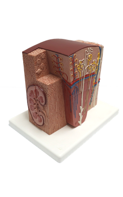
Main Model
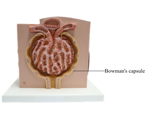
Zone F : Bowman's capsule
Urinary System
The urinary system consists of the two kidneys, the two ureters, the urinary bladder, and the urethra. This system regulates water and electrolyte homeostasis by the production of urine, the medium in which various metabolic waste products (particularly nitrogenous compounds such as urea and creatine) are eliminated. Urine, produced in the kidneys, passes down the ureters to the urinary bladder where it is temporarily stored and then eventually evacuated to the exterior through the urethra.
Each kidney is a large compound tubular gland and contains numerous nephrons. The nephron is the basic structural and functional unit; it consists of a renal corpuscle and a long, convoluted renal tubule. Nephrons are involved in (1) the formation of filtrate by ultrafiltration of blood plasma, (2) the selective resorption of most of the filtered water and other small molecules (such as amino acids, glucose, and sodium ions), and (3) the secretion of some excretory products (such as hydrogen and potassium ions). In addition, nephrons produce the hormones renin and erythropoietin. Renin is associated with the regulation of blood pressure, and erythropoietin stimulates production of erythrocytes in bone marrow.
The ureter is an excretory duct that drains urine away from the renal pelvis to the urinary bladder. Each ureter is lined by transitional epithelium. The urinary bladder is a hollow muscular organ, and is characterized by a thick lining of transitional epithelium. The urethra delivers urine from the urinary bladder to the outside. The male urethra is a duct common to the urinary and reproductive systems; the female urethra belongs only to the urinary system.
Kidney: Renal Pyramid
The kidneys are bean-shaped organs, 10-12 cm long and 3.5-5 cm thick. They are bilaterally positioned in the upper-posterior abdominal wall with one on either side of the vertebral column. Each kidney is invested by a tough fibroconnective tissue capsule that does not penetrate the parenchyma. The hilum of the kidney is a depressed region with a great deal of adipose tissue. Through this area the renal artery and vein, ureters, lymphatic vessels, and nerves enter or leave the kidney.
The basic structural and functional unit of the kidney is the nephron. It is responsible for filtration, excretion, and resorption. There are more than a million nephrons densely packed in each kidney. Each nephron consists of a renal corpuscle and a long, convoluted renal tubule, which is divided into a proximal convoluted tubule, loop of Henle, and a distal convoluted tubule that joins to the collecting tubule.
The substance of the kidney is made up of an outer cortex and an inner medulla. A portion of each nephron may be located in both cortex and medulla, but the renal corpuscles are only present in the cortex. The cortex can be divided into an outer zone and inner zone based on the distribution of the corpuscles. The medullary rays are groups of straight collecting tubules and loops of Henle in the cortex. Between the medullary rays are cortical labyrinths, composed of renal corpuscles and convoluted tubules. According to the combination of renal tubules at different areas, the medulla may also be divided into an outer zone and inner zone. The outer zone is further subdivided into an outer stripe and inner stripe.
The medulla is arranged into 10-18 pyramidal structures, known as renal pyramids. The base and sides of the renal pyramids are within the cortex, and their vertices, the renal papilla, protrude into the minor calyx. The renal pyramids are separated by a layer of connective tissue and the renal column, which possesses cortical structure. One renal pyramid is also called a lobe. A renal lobule is composed of a single medullary ray and the cortical tissue surrounding it.
After entering the hilum, the renal artery ramifies to form interlobar arteries, which run between the medullary pyramids. At the corticomedullary junction, the interlobar arteries give rise to arcuate arteries, which run parallel to the surface of the kidney. The interlobular arteries, derived from the arcuate arteries, run between the medullary rays toward the capsule. The interlobular arteries branch into several afferent glomerular arterioles, which supply the capillaries of the glomeruli. From there blood passes into the efferent glomerular arterioles, which branch again to form a second capillary network, supplying the majority of other portions of the same nephrons. The blood is collected into the interlobular veins, which join the arcuate veins. Then blood flows into the interlobar veins, which collectively form the renal vein through which blood leaves the kidney.
Cortex: Renal Corpuscle
The renal corpuscle, also known as a Malpighian corpuscle, is the essential portion of the nephron. It consists of a renal glomerulus and Bowman's capsule. The corpuscle has two poles: the vascular pole and urinary pole. At the vascular pole, the afferent arteriole (which supplies the capillary network of the glomerulus) enters, and the efferent arteriole (which collects the blood from the capillaries) leaves the corpuscle. The urinary pole is the beginning of the proximal convoluted tubule; it is continuous with Bowman's space.
The renal glomerulus is composed of a cluster of capillaries. After entering the corpuscle from the vascular pole, the afferent arteriole immediately branches into numerous capillaries that anastomose with one another and form a complicated capillary network. All capillaries eventually converge to form the efferent arteriole. The glomerulus is suspended in Bowman's capsule by means of the vascular pole. The endothelial cells of the capillary are fenestrated, rest on a thin basement membrane, and are externally surrounded by a layer of pedicels of the podocytes that constitute the visceral lining of Bowman's capsule. These three layers are called the glomerular filtration barrier, through which the filtrate first enters Bowman's space from the blood circulation. The capillary loops are supported by stalks of special connective tissue containing extraglomerular mesangial cells at the vascular pole. This cell type (mesangial cells) may also be found between the capillaries within the glomerulus.
Bowman's capsule is a double-walled envelope that holds the glomerulus. The parietal layer of the capsule is composed of simple squamous epithelial cells that are continuous with the cuboidal cells of the proximal tubules at the urinary pole. The visceral layer is made up of podocytes whose feet surround the glomerular capillaries, forming one of the layers of the filtration barrier.
The walls of afferent and efferent arterioles are composed of three layers, similar to those of arterioles elsewhere. At the vascular pole, however, the smooth muscle cells in the tunica media of the afferent arteriole are modified into epithelioid cells with a large nucleus and pale-staining cytoplasm. These are the juxtaglomerular cells, responsible for the synthesis of the hormone renin, which is associated with enhancing blood pressure.
The proximal convoluted tubule, starting at the urinary pole, is about 14 mm long and 50-60 micrometer wide. It follows a tortuous course through the cortical labyrinth, medullary ray, and the outer stripe of outer medulla, and is continuous with the thin segment of the loop of Henle. The proximal convoluted tubule is lined by a layer of cuboidal cells, which are characterized by the brush border at the free surface, striations at the basal surface, and a large, round nucleus in the center of the cytoplasm. The striations are recognized at the electron microscopic level as numerous mitochondria and an infolded basal cytoplasmic membrane. The function of these epithelial cells is to reabsorb the glucose, amino acids, ascorbic acids, chloride, and sodium from the filtrate within the tubule.
The distal convoluted tubule is situated between the straight part of the proximal tubule and the collecting tubule, and is composed of a straight part and a distal convoluted part. The epithelial cells lining the distal convoluted tubule are cuboidal, with a round nucleus and basal striations at the base of the cells. The distal convoluted tubule is responsible for the resorption of sodium, an activity that is promoted by the hormone aldosterone. At the portion of the distal convoluted tubule contacting the parent renal corpuscle between afferent and efferent arterioles, the epithelial cells become densely packed, with the nuclei close together. Thus the region appears darker and is known as macula densa. The macula cells are immediately adjacent to the afferent and efferent arterioles, to the juxtaglomerular cells, and to the extraglomerular mesangial cells. These three cell groups together are called the juxtaglomerular apparatus.
In the cortex, the connective tissue is sparse, with some collagen fibers and a small number of fibroblasts. However, there is an abundance of capillaries, which are supplied by the efferent arterioles.
Cortex: Medullary Ray
The medullary rays are finely striated extensions of the medullary tissue that project up into the cortex. They run parallel to the axis of the renal pyramid and represent the central axis of a renal lobule. A medullary ray consists of the straight portion of proximal tubules, the thick ascending limb of Henle's loops (distal tubules), and straight collecting tubules. The space between the tubules is filled with a very thin layer of loose connective tissue, containing some fibroblasts and a large number of capillaries.
Medulla: Outer Stripe of Outer Zone
The structure of the thick descending limb is similar to that of the proximal convoluted tubule. The thin descending limb may occasionally be found at the level of the medulla. It is lined by a simple squamous epithelium like a capillary. It differs from the capillary in several ways. For instance, the thin descending limb has a regular, rounded shape, its epithelial cells have thicker cytoplasm, and it lacks blood cells within the lumen. The thick ascending limb is lined by a layer of low cuboidal epithelial cells that resemble the distal convoluted tubule. These epithelial cells, however, are shorter and their round nuclei tend to bulge into the lumen. Additionally, they have a few microvilli at the free apical surface and striations at the base of the cell. These two parts of Henle's loop are involved in the resorption of water and sodium. The collecting tubule is lined by low cuboidal to tall columnar epithelial cells. The cells are generally pale-staining with a clear, regular cell border. Their nuclei are round or ovoid and situated at the center of the cell. The collecting tubules conduct urine from the nephron to the minor calyx, with some resorption of water.
The capillary network embedded in the sparse loose connective tissue containing a few fibroblasts can also be seen.
Medulla: Inner Stripe of Outer Zone
At this level, the thick descending limb (proximal tubule) is typically not present, but the thin descending limbs are quite abundant. Note that the connective tissue, containing fibroblasts and capillaries, gradually increases in volume.
Medulla: Inner Zone
At the inner medulla near the renal papilla, the collecting tubules converge to form papillary ducts, which convey urine into the pelvicalyceal space. The papillary ducts are lined by a layer of columnar or high columnar epithelial cells; they have a structure similar to that lining the collecting tubules. In addition to the papillary ducts, only thin segments of Henle's loop approach this inner zone. They run down to this zone and return to the outer medulla. There are no histological differences between the descending and ascending thin segment of Henle's loop.
Among these ducts and tubules there is a great amount of connective tissue. It contains capillaries, collagen fibers, and interstitial cells. These are fibroblast-like cells, whose function, apart from their collagen production, remains unknown.
The peripheral margin of the inner zone close to the papilla is covered by transitional epithelium, which is composed of only two or three layers of epithelial cells. These cells have a large nucleus, pale-staining cytoplasm, and a clear intercellular border.