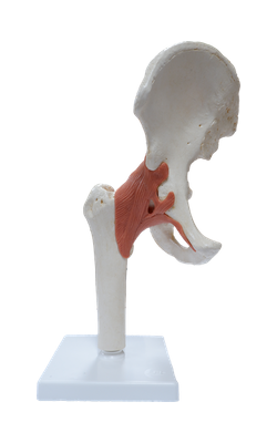
Main Model
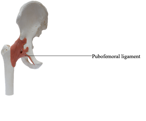
Pubofemoral ligament
Joint Capsule of Hip Joint
The hip joints are enclosed within strong joint capsules, formed of a loose external fibrous layer (fibrous capsule) and an internal synovial membrane. Proximally, the fibrous layer attaches to the acetabulum, just peripheral to the acetabular rim to which the labrum is attached, and to the transverse acetabular ligament. Distally, the fibrous layer attaches to the femoral neck only anteriorly at the intertrochanteric line and root of the greater trochanter. Posteriorly, the fibrous layer crosses the neck proximal to the intertrochanteric crest but is not attached to it.
Most fibers of the fibrous layer of the capsule take a spiral course from the hip bone to the intertrochanteric line of the femur, but some deep fibers pass circularly around the neck, forming the orbicular zone. Thick parts of the fibrous layer form the ligaments of the hip joint, which pass in a spiral fashion from the pelvis to the femur. Extension winds the spiraling ligaments and fibers more tightly, constricting the capsule and drawing the femoral head tightly into the acetabulum. The tightened fibrous layer increases the stability of the joint, but restricts extension of the joint to 10-20° beyond the vertical position. Flexion increasingly unwinds the spiraling ligaments and fibers. This permits considerable flexion of the hip joint with increasing mobility.
Of the three intrinsic ligaments of the joint capsule below, it is the first one that reinforces and strengthens the joint:
• Anteriorly and superiorly is the strong, Y-shaped iliofemoral ligament, which attaches to the anterior inferior iliac spine and the acetabular rim proximally and the intertrochanteric line distally. Said to be the body's strongest ligament, the iliofemoral ligament specifically prevents hyperextension of the hip joint during standing by screwing the femoral head into the acetabulum.
• Anteriorly and inferiorly is the pubofemoral ligament, which arises from the obturator crest of the pubic bone and passes laterally and inferiorly to merge with the fibrous layer of the joint capsule. This ligament blends with the medial part of the iliofemoral ligament, and tightens during both extension and abduction of the hip joint. The pubofemoral ligament prevents overabduction of the hip joint.
• Posteriorly is the ischiofemoral ligament, which arises from the ischial part of the acetabular rim. The weakest of the three ligaments, it spirals superolaterally to the femoral neck, medial to the base of the greater trochanter.
The ligaments and peri-articular muscles (the medial and lateral rotators of the thigh) play a vital role in maintaining the structural integrity of the joint.