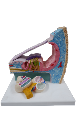
Main Model
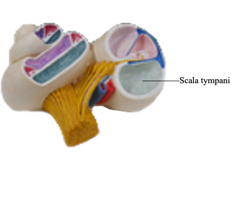
General view of the cochlear : Scala tympani
Membranous Labyrinth
The membranous labyrinth consists of a series of communicating sacs and ducts that are suspended in the bony labyrinth. The labyrinth contains endolymph, a watery fluid similar in composition to intracellular fluid, thus differing in composition from the surrounding perilymph (which is like extracellular fluid) that fills the remainder of the bony labyrinth. The membranous labyrinth - composed of two divisions, the vestibular labyrinth and the cochlear labyrinth - consists of more parts than does the bony labyrinth:
• Vestibular labyrinth: utricle and saccule, two small communicating sacs in the vestibule of the bony labyrinth.
• Three semicircular ducts in the semicircular canals.
• Cochlear labyrinth: cochlear duct in the cochlea.
The spiral ligament, a spiral thickening of the periosteal lining of the cochlear canal, secures the cochlear duct to the spiral canal of the cochlea. The remainder of the membranous labyrinth is suspended by delicate filaments that traverse the perilymph.
The semicircular ducts open into the utricle through five openings, reflective of the way the surrounding semicircular canals open into the vestibule. The utricle communicates with the saccule through the utriculosaccular duct, from which the endolymphatic duct arises. The saccule is continuous with the cochlear duct through the ductus reuniens, a uniting duct. The utricle and saccule have specialized areas of sensory epithelium called maculae. The macula of the utricle (Latin macula utriculi) is in the floor of the utricle, parallel with the base of the cranium, whereas the macula of the saccule (Latin macula sacculi) is vertically placed on the medial wall of the saccule. The hair cells in the maculae are innervated by fibers of the vestibular division of the vestibulocochlear nerve. The primary sensory neurons are in the vestibular ganglia, which are in the internal acoustic meatus.
The endolymphatic duct traverses the vestibular aqueduct and emerges through the bone of the posterior cranial fossa, where it expands into a blind pouch called the endolymphatic sac. The endolymphatic sac is located under the dura mater on the posterior surface of the petrous part of the temporal bone. The sac is a storage reservoir for excess endolymph, formed by the blood capillaries in the membranous labyrinth.
Semicircular Ducts
Each semicircular duct has an ampulla at one end containing a sensory area, the ampullary crest (Latin crista ampullari). The crests are sensors for recording movements of the endolymph in the ampulla resulting from rotation of the head in the plane of the duct. The hair cells of the crests, like those of the maculae, stimulate primary sensory neurons, whose cell bodies are in the vestibular ganglia.
Cochlear Duct
The cochlear duct is a spiral tube, closed at one end and triangular in cross section. The duct is firmly suspended across the cochlear canal between the spiral ligament on the external wall of the cochlear canal and the osseous spiral lamina of the modiolus. Spanning the spiral canal in this manner, the endolymph-filled cochlear duct divides the perilymph-filled spiral canal into two channels that are continuous at the apex of the cochlea at the helicotrema, a semilunar communication at the apex of the cochlea.
Waves of hydraulic pressure created in the perilymph of the vestibule by the vibrations of the base of the stapes ascend to the apex of the cochlea by one channel, the scala vestibuli. The pressure waves then pass through the helicotrema and descend back to the basal turn of the cochlea by the other channel, the scala tympani. Here, the pressure waves again become vibrations, this time of the secondary tympanic membrane in the round window, and the energy initially received by the (primary) tympanic membrane is finally dissipated into the air of the tympanic cavity.
The roof of the cochlear duct is formed by the vestibular membrane. The floor of the duct is also formed by part of the duct, the basilar membrane, plus the outer edge of the osseous spiral lamina. The receptor of auditory stimuli is the spiral organ (of Corti), situated on the basilar membrane. It is overlaid by the gelatinous tectorial membrane.
The spiral organ contains hair cells, the tips of which are embedded in the tectorial membrane. The organ is stimulated to respond by deformation of the cochlear duct induced by the hydraulic pressure waves in the perilymph, which ascend and descend in the surrounding scalae vestibuli and tympani.