Main Model
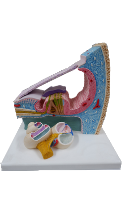
Organ of Corti : Tectorial membrane
Inner Ear: Structure of the Cochlea
The cochlea is named for its similarity to a conch shell. Its main elements include the fluid-filled membranous labyrinth, specialized sensory epithelium of the organ of Corti, and neurons of the spiral ganglion with their peripheral and central axonal branches.
The membranous cochlea, the coiled portion of the inner ear, is encased in the osseous cochlea and consists of three spiraling chambers. The cochlea makes approximately two and two-thirds turns from base to apex. Uncoiled, it is about 34 mm long. The base of the cochlear spiral is connected to the saccule of the membranous labyrinth by the ductus reuniens.
The central chamber of the membranous cochlea is the cochlear duct, also called the scala media. Above it, the scala vestibuli is positioned to communicate with the vestibule, the portion of the membranous inner ear between the oval window and the cochlea. Below, the scala tympani ends at the round window, which separates this space from the middle ear cavity. In cross section, the scala media is bounded by the basilar membrane below, the vestibular or Reissner membrane above, and the stria vascularis externally. The screw-like bony core of the cochlea is the modiolus. A spiral osseous lamina extends outward from the modiolus to join the basilar membrane. The basilar membrane, in turn, is continuous laterally with the spiral ligament. The scala vestibuli and tympani are filled with perilymph. The endolymph, which fills the cochlear duct, is elaborated by the cells and rich capillary bed of the stria vascularis.
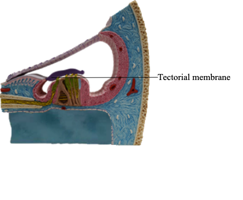
The organ of Corti is the specialized sensory epithelium resting on the basilar membrane. It is composed of inner and outer hair cells, supporting cells, and the tectorial membrane. The inner hair cells are separated from the outer hair cells by the tunnel of Corti. This tunnel is formed by the filamentous arches of the inner and outer pillar cells and is filled with fluid.
Inner hair cells form a single line spiraling from base to apex, and the outer hair cells form three parallel lines that follow the same course. Once damaged, human hair cells do not regenerate. Research has not yet found a way to augment this. It is uncertain how many of the inner (about 3500) or outer (about 12,000) hair cells must be lost to disease, trauma, or aging before a just-noticeable sensorineural hearing loss ensues. Projecting from the apical surface of each hair cell is a hair bundle consisting of 50 to 150 stereocilia arranged in curving rows. Each hair bundle is polarized so that the longest stereocilia are on the outer border, and the rows of stereocilia are linked by filamentous material at their tips.
The tectorial membrane is a gelatinous arm that extends outward over the sensory epithelium from the limbus of the osseous spiral lamina. The taller stereocilia in each hair bundle are in contact with or embedded in the tectorial membrane. Consequently, movement of the basilar membrane and the organ of Corti will bend the stereocilia against the tectorial membrane and cause a graded depolarization of the hair cells.
The bony modiolus, around which the cochlear duct turns, houses the spiral ganglion. At the edge of the osseous spiral lamina, the peripheral processes of the bipolar cells of this ganglion lose their myelin and pass through perforations to the basilar membrane, where they synapse on the base of the inner and outer hair cells. The central processes of the spiral ganglion cells form the cochlear portion of the vestibulocochlear nerve (cranial nerve VIII). Efferent fibers to the cochlea either spiral along the inner part of the basilar membrane to synapse on inner hair cells or travel radially across the tunnel of Corti to contact outer hair cells.