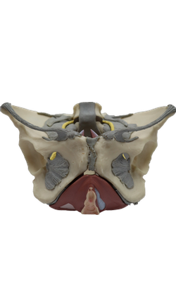
Main Model
MUSCLES : Levator ani + Pelvic diaphragm
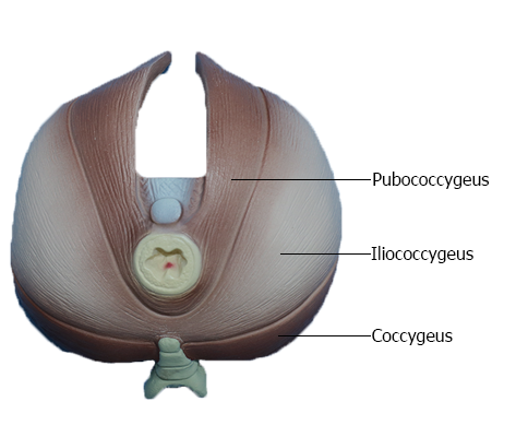
Pelvic Floor
The pelvic floor is formed by the bowl- or funnel-shaped pelvic diaphragm, which consists of the coccygeus and levator
ani muscles and the fascias (Latin fasciae) covering the superior
and inferior aspects of these muscles. The pelvic diaphragm lies within the lesser
pelvis, separating the pelvic cavity from the perineum, for
which it forms the roof.
The attachment of the diaphragm to the obturator fascia
divides the obturator internus into a superior pelvic portion
and an inferior perineal portion. Medial to the
pelvic portions of the obturator internus muscles are the
obturator nerves and vessels and other branches of the internal iliac vessels.
The coccygeus muscles arise from the lateral aspects of
the inferior sacrum and coccyx, their fleshy fibers lying on and
attaching to the deep surface of the sacrospinous ligament. The levator ani (a broad muscular sheet)
is the larger and more important part of the pelvic floor. It is
attached to the bodies of the pubic bones anteriorly, to the ischial spines posteriorly, and to a thickening in the obturator
fascia (the tendinous arch of the levator ani) between the two bony sites on each side.
The pelvic diaphragm thus stretches between the anterior, the lateral, and the posterior walls of the lesser pelvis, giving it the appearance of a hammock suspended from these
attachments, closing much of the ring of the pelvic girdle. An
anterior gap between the medial borders of the levator ani
muscles of each side - the urogenital hiatus - gives passage to the urethra and, in females, the vagina.
The levator ani consists of three parts, often poorly
demarcated but designated according to attachments and
fiber course:
• Puborectalis: the thicker, narrower, medial part of the
levator ani, consisting of muscle fibers that are continuous between the posterior aspects of the bodies of the
right and left pubic bones. It forms a U-shaped muscular sling (puborectal sling) that passes posterior to the anorectal junction, bounding the urogenital
hiatus. This part plays a major role in maintaining fecal
continence.
• Pubococcygeus: the wider but thinner intermediate part
of the levator ani, which arises lateral to the puborectalis from the posterior aspect of the body of the pubis and
anterior tendinous arch.
It passes posteriorly in a nearly horizontal plane; its lateral
fibers attach to the coccyx and its medial fibers merge with
those of the contralateral muscle to form a fibrous raphe
or tendinous plate, part of the anococcygeal body or
ligament between the anus and the coccyx (often referred
to clinically as the "levator plate").
Shorter muscular slips of the pubococcygeus extending medially and blending with the fascia around midline
structures are named for the structure near their termination: pubovaginalis (females), puboprostaticus (males),
puboperinealis, and pubo-analis.
• Iliococcygeus: the posterolateral part of the levator ani,
which arises from the posterior tendinous arch and ischial
spine. It is thin and often poorly developed (appearing
more aponeurotic than muscular), and also blends with
the anococcygeal body posteriorly.
The levator ani forms a dynamic floor for supporting the abdominopelvic viscera. It is tonically contracted most of the time
to support the abdominopelvic viscera, and to assist in maintaining urinary and fecal continence. It is actively contracted
during activities such as forced expiration, coughing, sneezing,
vomiting, and fixation of the trunk during strong movements
of the upper limbs (e.g., when lifting heavy objects), primarily
to increase support of the viscera during periods of increased
intra-abdominal pressure, and perhaps secondarily to contribute to the increased pressure (to aid expulsion).
Penetrated centrally by the anal canal, the levator ani
is funnel shaped, with the U-shaped puborectalis looping
around the "funnel spout"; its tonic contraction bends the
anorectum anteriorly. Active contraction of the (voluntary)
puborectalis portion is important in maintaining fecal continence immediately after rectal filling or during peristalsis
when the rectum is full and the involuntary sphincter muscle
is inhibited (relaxed).
The levator ani must relax to allow urination and defecation. The increased intra-abdominal pressure for defecation
is provided by contraction of the (thoracic) diaphragm and
muscles of the anterolateral abdominal wall. Acting together,
the parts of the levator ani elevate the pelvic floor after their
relaxation and the consequent descent of the pelvic diaphragm that occurs during urination and defecation.
NB: Muscles of Floor of Pelvis
Pelvic diaphragm = Levator ani + Coccygeus
Levator ani = Pubococcygeus + Iliococcygeus
Pubococcygeus (Female) = Puborectalis + Pubovaginalis
Pubococcygeus (Male) = Puborectalis + Puboprostaticus (Levator prostatae)