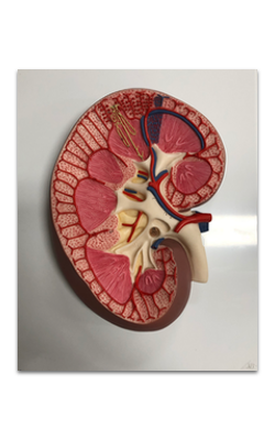
Main Model
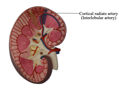
7 Cortical radiate artery (Interlobular artery)
Kidney: Renal Pyramid
The kidneys are bean-shaped organs, 10-12 cm long and 3.5-5 cm thick. They are bilaterally positioned in the upper-posterior abdominal wall with one on either side of the vertebral column. Each kidney is invested by a tough fibroconnective tissue capsule that does not penetrate the parenchyma. The hilum of the kidney is a depressed region with a great deal of adipose tissue. Through this area the renal artery and vein, ureters, lymphatic vessels, and nerves enter or leave the kidney.
The basic structural and functional unit of the kidney is the nephron. It is responsible for filtration, excretion, and resorption. There are more than a million nephrons densely packed in each kidney. Each nephron consists of a renal corpuscle and a long, convoluted renal tubule, which is divided into a proximal convoluted tubule, loop of Henle, and a distal convoluted tubule that joins to the collecting tubule.
The substance of the kidney is made up of an outer cortex and an inner medulla. A portion of each nephron may be located in both cortex and medulla, but the renal corpuscles are only present in the cortex. The cortex can be divided into an outer zone and inner zone based on the distribution of the corpuscles. The medullary rays are groups of straight collecting tubules and loops of Henle in the cortex. Between the medullary rays are cortical labyrinths, composed of renal corpuscles and convoluted tubules. According to the combination of renal tubules at different areas, the medulla may also be divided into an outer zone and inner zone. The outer zone is further subdivided into an outer stripe and inner stripe.
The medulla is arranged into 10-18 pyramidal structures, known as renal pyramids. The base and sides of the renal pyramids are within the cortex, and their vertices, the renal papilla, protrude into the minor calyx. The renal pyramids are separated by a layer of connective tissue and the renal column, which possesses cortical structure. One renal pyramid is also called a lobe. A renal lobule is composed of a single medullary ray and the cortical tissue surrounding it.
After entering the hilum, the renal artery ramifies to form interlobar arteries, which run between the medullary pyramids. At the corticomedullary junction, the interlobar arteries give rise to arcuate arteries, which run parallel to the surface of the kidney. The interlobular arteries, derived from the arcuate arteries, run between the medullary rays toward the capsule. The interlobular arteries branch into several afferent glomerular arterioles, which supply the capillaries of the glomeruli. From there blood passes into the efferent glomerular arterioles, which branch again to form a second capillary network, supplying the majority of other portions of the same nephrons. The blood is collected into the interlobular veins, which join the arcuate veins. Then blood flows into the interlobar veins, which collectively form the renal vein through which blood leaves the kidney.