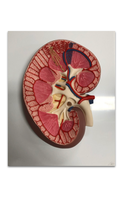
Main Model
19 Renal corpuscle
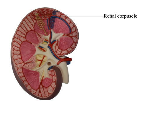
The Renal Corpuscle
The renal corpuscle, or malpighian corpuscle, consists of the capsule of Bowman
investing a capillary tuft, the glomerulus.
The capsule of Bowman has two layers:
1. The visceral layer, attached to the capillary
glomerulus.
2. The parietal layer, facing the connective tissue
stroma.
The visceral layer is lined by epithelial cells called
podocytes supported by a basal lamina. The parietal
layer consists of a simple squamous epithelium continuous with the simple cuboidal epithelium of the
proximal convoluted tubule.
A urinary space (Bowman's space or capsular
space), containing the plasma ultrafiltrate (primary
urine), exists between the visceral and parietal layers
of the capsule.
The urinary space is continuous with the lumen of
the proximal convoluted tubule at the urinary pole,
the gate through which the plasma ultrafiltrate flows
into the proximal convoluted tubule. The opposite pole, the site of entry and exit of the afferent and efferent glomerular arterioles, is called the vascular pole.
The glomerulus consists of three cell components:
1. The podocytes, the visceral layer of the capsule
of Bowman.
2. The fenestrated endothelial cells, lining the
glomerular capillaries.
3. The mesangial cells, embedded in the mesangial
matrix. Mesangium designates the combined mesangial cells-mesangial matrix complex.