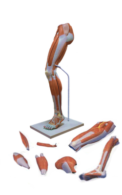
Main Model

21 Femoral artery

The femoral triangle, a subfascial formation, is a
triangular landmark useful in dissection and in understanding
relationships in the groin. In living people, it appears as a triangular
depression inferior to the inguinal ligament when the thigh is flexed,
abducted, and laterally rotated.
The contents of the femoral triangle, from lateral to medial, are the:
• Femoral nerve and its (terminal) branches.
• Femoral sheath and its contents:
- Femoral artery and several of its branches.
- Femoral vein and its proximal tributaries (e.g., the great saphenous and profunda femoris veins).
- Deep inguinal lymph nodes and associated lymphatic vessels.
• Femoral nerve and its (terminal) branches.
• Femoral sheath and its contents:
- Femoral artery and several of its branches.
- Femoral vein and its proximal tributaries (e.g., the great saphenous and profunda femoris veins).
- Deep inguinal lymph nodes and associated lymphatic vessels.
The femoral triangle is bisected by the femoral
artery and vein, which pass to and from the adductor canal inferiorly at
the triangle’s apex. The adductor canal is an intermuscular passageway
deep to the sartorius by which the major neurovascular bundle of the
thigh traverses the middle third of the thigh.