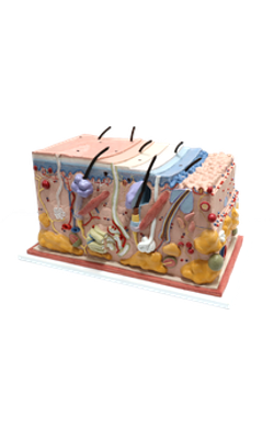
Main Model

Anterior : Sebaceous gland

Sebaceous Gland
The sebaceous glands are distributed throughout the skin, except in the palms and the soles. Generally their ducts are associated with hair follicles, but those of the eyelid tarsal glands, lip, glans penis, inner surface of the prepuce, and labia minora open directly onto the skin surface.
Sebaceous glands are located in the dermis, surrounded by a thin layer of connective tissue. These are saccular glands, with a short duct that opens into the space, which lies between the external root sheath of the hair follicle and the cortex of the hair. The secretory acini are supported by a delicate basement membrane. The basal cells, which rest on the basement membrane, are small cuboidal cells and form a single layer continuous with the basal cells of the dermis. Toward the center of the acinus, they become larger secretory cells, which possess clear foamlike cytoplasm filled with lipid droplets, and the nuclei gradually shrink and then disappear. Near the duct, secretory cells degenerate and break down into a fatty mass and cellular debris; thus, the entire cell becomes secretion, sebum. This process is known as holocrine secretion. The sebum is an oily secretion, containing lipids, triglycerides, squalene, wax esters, and sterol. It lubricates skin and hairs. Cells lost in the secretory process are replaced by division of the basal cells of the acini. Discharge of secretion is aided by contraction of the arrector pili muscle. It is attached at one end to the middle of the connective tissue sheath of the follicle and at the other end to the papillary layers of the dermis.