Main Model
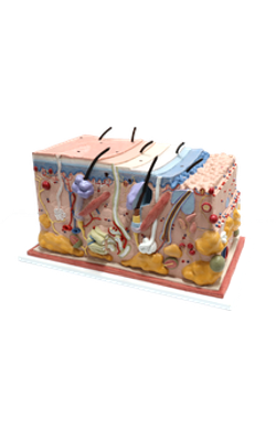
Anterior : Medulla of hair

Epidermal Derivatives: Hair (Pilosebaceous Unit)
Scattered in the epidermis are the hair follicles. During development, the epidermis and dermis interact to develop sweat glands and hair follicles.
A hair follicle primordium (called the hair germ) forms as a cell aggregate in the basal layer of the epidermis, induced by signaling molecules derived from fibroblasts of the dermal mesoderm. As basal epidermal cell clusters extend into the dermis, dermal fibroblasts form a small nodule (called a dermal papilla) under the hair germ.
The dermal papilla pushes into the core of the hair germ, whose cells divide and differentiate to form the keratinized hair shaft. Melanocytes present in the hair germ produce and transfer melanin into the shaft.
A bulbous swelling (called the follicular bulge) on the side of the hair germ contains stem cells, clonogenic keratinocytes, that can migrate and regenerate the hair shaft, the epidermis, and sebaceous gland, forming pilosebaceous units, in response to morphogenetic signals.
The first adult hair follicle cycle starts once morphogenesis is completed about 18 days after birth, The first hair in the human embryo is thin, unpigmented, and spaced, and is called lanugo. Lanugo is shed before birth and replaced by short colorless hair called vellus. Terminal hair replaces vellus, which remains in the so-called hairless parts of the skin (such as the forehead of the adult and armpits of infants).
Hair follicles are tubular invaginations of the epidermis responsible for the growth of hair.
Hair follicles are constantly cycling between:
1. Growth (anagen) phase.
2. Regression (catagen) phase.
3. Resting (telogen) phase.
During the first 28 days of the telogen phase, hair follicles become quiescent because of growth inhibitory signals from the dermis (mainly from bone morphogenetic proteins). Increased Wnt/beta-catenin signaling promotes stem cell activation to initiate the growth of new hair during the transition from telogen to anagen. Anagen, catagen and telogen will sequentially continue during the life of the individual.
Each hair follicle consists of two parts:
1. The hair shaft.
2. The hair bulb.
The hair shaft is a filamentous keratinized structure present almost all over the body surface, except on the thick skin of the palms and soles, the sides of the fingers and toes, the nipples, and the glans penis and the clitoris, among others.
A cross section of the hair shaft of thick hair reveals three concentric zones containing keratinized cells:
1. The cuticle.
2. The cortex.
3. The medulla (the last is absent in thin hair).
The hair shaft consists of hard keratin.
The hair bulb is the expanded end portion of the invaginated hair follicle. A vascularized connective tissue core (dermal papilla) projects into the hair bulb, in close proximity to matrix cells.
The hair shaft is surrounded by:
1. The external root sheath, a downgrowth of the epidermis.
2. The internal root sheath, generated by the hair bulb (the hair matrix cells), is made up of three layers of soft keratin (which from the outside to the inside are the Henle's layer, the Huxley's layer, and the cuticle of the inner root sheath, adjacent to the cuticle of the hair shaft).
The keratinization of the hair and internal root sheath occurs in a region called the keratogenous zone, the transition zone between maturing epidermal cells and hard keratin. The external root sheath is not derived from the hair bulb.
The hair follicle is surrounded by a connective tissue layer and associated with the arrector pili muscle, a bundle of smooth muscle fibers aligned at an oblique angle to the connective tissue sheath and the epidermis. The autonomic nervous system controls the arrector pili muscle, which contracts during fear, strong emotions, and cold temperature. The hairs stand up and the attachment site of the muscle bundle at the epidermis forms a small groove, the so called goose flesh.
The hair follicle is associated with sebaceous glands with their excretory duct connected to the lumen of the hair follicle. When the arrector pili muscle contracts and the hair stands up, sebum is forced out of the sebaceous gland into the lumen of the hair follicle.
The color of the hair depends on the amount and distribution of melanin in the hair shaft. Few melanosomes are seen in blond hair. Few melanocytes and melanin are seen in gray hair. Red hair has a chemically distinct melanin, and melanosomes are round rather than ellipsoid.
A structure that is not recognized in routine histologic sections of hairs is the peritrichial nerve endings wrapped around the base of the hair follicle. The nerve is stimulated by hair movement.