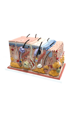
Main Model
Anterior : Melanocyte

Melanocytes
Melanocytes are branching cells located in the stratum basale of the epidermis. Melanocytes derive from melanoblasts, a cell
precursor migrating from the neural crest.
The development of the melanoblast into melanocytes is under the control of the ligand stem cell factor
interacting with the c-kit receptor, a membrane-bound tyrosine kinase.
The development of mast cells, primordial germinal
cells, and hematopoietic stem cells is also dependent on the interaction of stem cell factor with the c-kit
receptor.
Melanocytes enter the developing epidermis and
remain as independent cells without desmosome
attachment to the differentiating keratinocytes.
The turnover of melanocytes is slower than that of
keratinocytes.
Melanocytes produce melanin, contained in
melanosomes, which are transferred to neighboring keratinocytes through their branching cell processes,
called melanocyte dendrites, and released by cytocrine secretion.
Melanins are pigments that provide the skin
and hairs (by cell transfer) and eyes (for storage in pigmented epithelia of the retina and ciliary body
and iris) with color and photoprotection against ionizing radiation. Melanins consist of copolymers
of black and brown eumelanins and red and yellow
pheomelanins.
Melanosomes develop and mature in melanocytes
through four distinct stages:
1. During the first and second stages, premelanosomes, derived from the early endosome compartment by a sorting mechanism driven by membrane-bound adaptor proteins-3 and -1 (AP-3 and AP-1),
contain PMEL fibrils but lack melanin pigment.
PMEL fibrils are cleaved to M alpha and M beta fragments
by the enzyme proprotein convertase. M alpha fragments begin to form melanofilaments, the scaffold
for melanin deposition. Protein AP-3-depending premelanosome sorting is defective in the genetic
disease Hermansky-Pudlack syndrome (HPS), characterized by oculocutaneous albinism, bleeding
caused by a deficiency or absence of platelet stored
granules and, in some cases, pulmonary fibrosis or
granulomatous colitis.
2. The third stage starts once the melanofilaments
are fully formed and the synthesis of melanin starts
within the premelanosome by the activity of melanin
biosynthetic enzymes tyrosinase, tyrosinase-related
protein-1 and DOPAchrome tautomerase, also
sorted as cargo from AP-3-coated endosomal buds
to premelanosomes.
Melanin is produced by oxidation of tyrosine to
3,4-dihydroxyphenylalanine (DOPA). Oxidation is
catalyzed by tyrosinase, whose activity is modulated
by tyrosinase-related protein-1. DOPA is then transformed to eumelanin, which accumulates on the pre-assembled M alpha-containing melanofilament scaffold.
3. The fourth stage is completed when the internal
fibrillar structure of the premelanosome is masked by
deposits of melanin and melanosomes are transported
along microtubules by the motor protein kinesin to actin-containing melanocyte dendritic tips to be
transferred to adjacent keratinocytes.
Melanosome transfer occurs when melanophilin,
an adapter protein, binds to Rab27a, a protein inserted in the melanosome membrane. The F-actin-based molecular motor myosin
Va binds to the Rab27a-melanophilin complex and
transports the melanosome to the plasma membrane. Extruded melanin by exocytosis is captured by adjacent keratinocytes and internalized by endocytosis.
Albinism results from the inability of cells to
form melanin. Griscelli syndrome is determined
by mutations of the myosin Va gene. Patients with
Griscelli syndrome have silvery hair, partial albinism,
occasional neurologic defects, and immunodeficiency
(due to a defective vesicular transport and secretion
in cytolytic T cells). Similar pigmentation disorders
are determined by mutations in the Rab27a and
melanophilin genes.