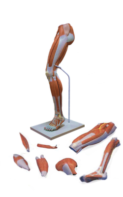
Main Model
13 Tibia (Shin bone)
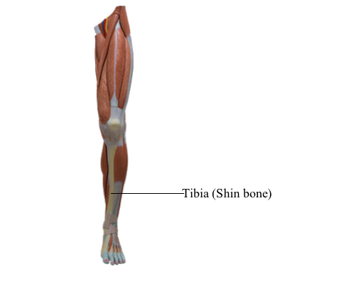
Located on the anteromedial side of the leg, nearly parallel to the fibula, the tibia (shin bone) is the second largest bone in the body. It flares outward at both ends to provide an increased area for articulation and weight transfer. The superior (proximal) end widens to form medial and lateral condyles that overhang the shaft medially, laterally, and posteriorly, forming a relatively flat superior articular surface, or tibial plateau. This plateau consists of two smooth articular surfaces (the medial one slightly concave and the lateral one slightly convex) that articulate with the large condyles of the femur. The articular surfaces are separated by an intercondylar eminence formed by two intercondylar tubercles (medial and lateral) flanked by relatively rough anterior and posterior intercondylar areas.
The tubercles fit into the intercondylar fossa between the femoral condyles. The intercondylar tubercles and areas provide attachment for the menisci and principal ligaments of the knee, which hold the femur and tibia together, maintaining contact between their articular surfaces.
The anterolateral aspect of the lateral tibial condyle bears an anterolateral tibial tubercle (Gerdy tubercle) inferior to the articular surface, which provides the distal attachment for a dense thickening of the fascia covering the lateral thigh, adding stability to the knee joint. The lateral condyle also bears a fibular articular facet posterolaterally on its inferior aspect for the head of the fibula.
Unlike that of the femur, the shaft of the tibia is truly vertical within the leg. It is somewhat triangular in cross-section, having three surfaces and borders: medial, lateral/ interosseous, and posterior.
The anterior border of the tibia is the most prominent border. It and the adjacent medial surface are subcutaneous throughout their lengths and are commonly known as the “shin”. Their periosteal covering and overlying skin are vulnerable to bruising. At the superior end of the anterior border, a broad, oblong tibial tuberosity provides distal attachment for the patellar ligament, which stretches between the inferior margin of the patella and the tibial tuberosity.
The tibial shaft is thinnest at the junction of its middle and distal thirds. The distal end of the tibia is smaller than the proximal end, flaring only medially; the medial expansion extends inferior to the rest of the shaft as the medial malleolus. The inferior surface of the shaft and the lateral surface of the medial malleolus articulate with the talus and are covered with articular cartilage.
The interosseous border of the tibia is sharp where it gives attachment to the interosseous membrane that unites the two leg bones. Inferiorly, the sharp border is replaced by a groove, the fibular notch, that accommodates and provides fibrous attachment to the distal end of the fibula.
On the posterior surface of the proximal part of the tibial shaft is a rough diagonal ridge, called the soleal line, which runs inferomedially to the medial border. This line is formed in relationship to the aponeurotic origin of the soleus muscle approximately one third of the way down the shaft. Immediately distal to the soleal line is an obliquely directed vascular groove, which leads to a large nutrient foramen for passage of the main artery supplying the proximal end of the bone and its marrow. From it, the nutrient canal runs inferiorly in the tibia before it opens into the medullary (marrow) cavity.