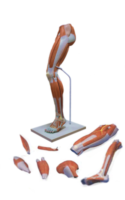
Main Model
29 Popliteal artery
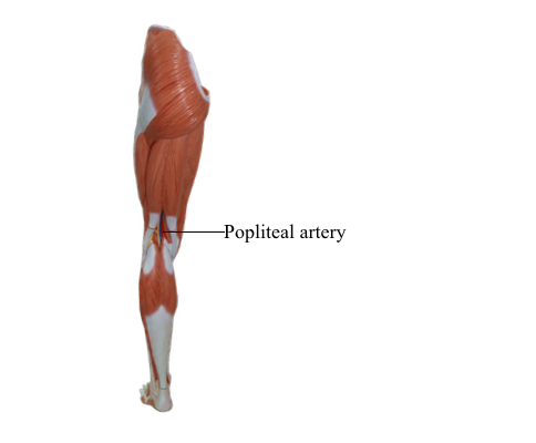
Blood Vessels in Popliteal Fossa
The popliteal artery, the continuation of the femoral artery, begins when the latter passes through the adductor hiatus. The popliteal artery passes inferolaterally through the fossa and ends at the inferior border of the popliteus by dividing into the anterior and posterior tibial arteries. The deepest (most anterior) structure in the fossa, the popliteal artery, runs in close proximity to the joint capsule of the knee as it spans the intercondylar fossa.
Five genicular branches of the popliteal artery supply the capsule and ligaments of the knee joint. The genicular arteries are the superior lateral, superior medial, middle, inferior lateral, and inferior medial genicular arteries. They participate in the formation of the peri-articular genicular anastomosis, a network of vessels surrounding the knee that provides collateral circulation capable of maintaining blood supply to the leg during full knee flexion, which may kink the popliteal artery. Other contributors to this important genicular anastomosis are the:
• Descending genicular artery, a branch of the femoral artery, superomedially.
• Descending branch of the lateral femoral circumflex artery, superolaterally.
• Anterior tibial recurrent artery, a branch of the anterior tibial artery, inferolaterally.
Muscular branches of the popliteal artery supply the hamstring, gastrocnemius, soleus, and plantaris muscles. The superior muscular branches of the popliteal artery have clinically important anastomoses with the terminal part of the profunda femoris and gluteal arteries.
The popliteal vein begins at the distal border of the popliteus as a continuation of the posterior tibial vein. Throughout its course, the vein lies close to the popliteal artery, lying superficial to it and in the same fibrous sheath. The popliteal vein is initially posteromedial to the artery and lateral to the tibial nerve. More superiorly, the popliteal vein lies posterior to the artery, between this vessel and the overlying tibial nerve. Superiorly, the popliteal vein, which has several valves, becomes the femoral vein as it traverses the adductor hiatus. The small saphenous vein passes from the posterior aspect of the lateral malleolus to the popliteal fossa, where it pierces the deep popliteal fascia and enters the popliteal vein.