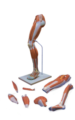
Main Model
49 Common fibular nerve (Common peroneal nerve)

Nerves in Popliteal Fossa
The sciatic nerve usually ends at the superior angle of the popliteal fossa by dividing into the tibial and common fibular nerves.
The tibial nerve is the medial, larger terminal branch of the sciatic nerve derived from anterior (preaxial) divisions of the anterior rami of the L4-S3 spinal nerves. The tibial nerve is the most superficial of the three main central components of the popliteal fossa (i.e., nerve, vein, and artery); however, it is still in a deep and protected position. The tibial nerve bisects the fossa as it passes from its superior to its inferior angle.
While in the fossa, the tibial nerve gives branches to the soleus, gastrocnemius, plantaris, and popliteus muscles. The medial sural cutaneous nerve is also derived from the tibial nerve in the popliteal fossa. It is joined by the sural communicating branch of the common fibular nerve at a highly variable level to form the sural nerve. This nerve supplies the lateral side of the leg and ankle.
The common fibular (peroneal) nerve is the lateral, smaller terminal branch of the sciatic nerve derived from posterior (post axial) divisions of the anterior rami of the L4-S2 spinal nerves. The common fibular nerve begins at the superior angle of the popliteal fossa and follows closely the medial border of the biceps femoris and its tendon along the superolateral boundary of the fossa. The nerve leaves the fossa by passing superficial to the lateral head of the gastrocnemius and then passes over the posterior aspect of the head of the fibula. The common fibular nerve winds around the neck of the fibula and divides into its terminal branches.
The most inferior branches of the posterior cutaneous nerve of the thigh supply the skin that overlies the popliteal fossa. The nerve traverses most of the length of the posterior compartment of the thigh deep to the fascia lata; only its terminal branches enter the subcutaneous tissue as cutaneous nerves.