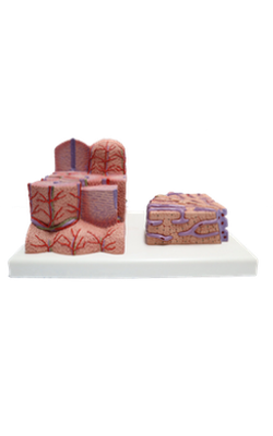
Main Model
Bile canaliculi : Superior
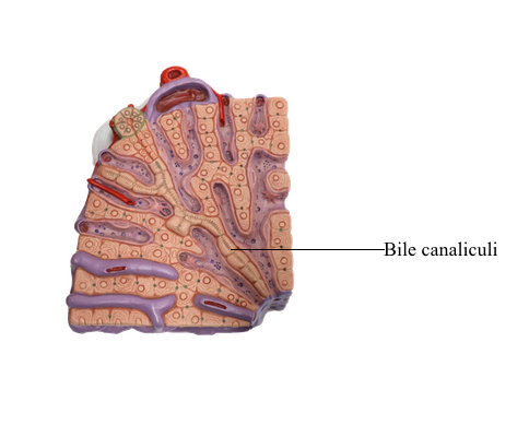
Bile canaliculi, about 0.5-1.0 micrometer in diameter, are formed by the cytoplasmic membranes of adjacent hepatocytes. They therefore have no wall of their own. The bile canaliculi are not easily seen in conventional preparations, but they are revealed by special staining methods, such as Golgi's silver impregnation. In the plane of the hepatic plate, each hepatocyte is surrounded by a hexagonal network of bile canaliculi. The canaliculi anastomose with one another to form a three-dimensional network. In the periphery of the lobule, they connect with the canal of Hering, which drains into the bile duct in the portal triad.
Bile contains bile salts, bilirubin, and steroids; bile salts are associated with the digestion and absorption of fats. The bile made by the hepatocytes is first secreted into the lumen of the bile canaliculi. When bile canaliculi are broken because of the necrosis of hepatocytes or obstruction of the bile duct, bile enters the blood stream through the space of Disse, resulting in the accumulation of bilirubin. Bilirubin is a metabolic product of hemoglobin and imparts a yellow appearance to the skin and sclera, a condition known as jaundice.