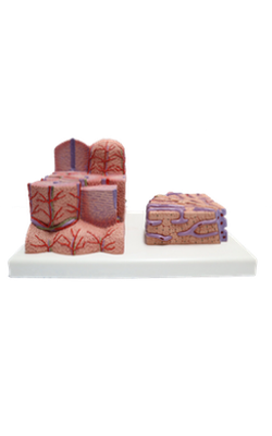
Main Model
Central vein : Superior View 2
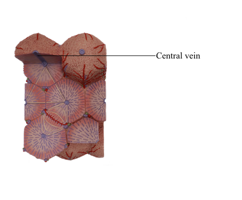
As a structural unit of the liver, each hepatic lobule has a central vein in the middle, with hepatic plates radiating from it to the periphery like the spokes of a wheel from a central hub. The hepatic plate consists of a single layer of hepatocytes, anastomosing with one another to form a network in three dimensions. Between the hepatic plates are sinusoids filled with blood and phagocytic cells. The human liver has a structural pattern of lobules similar to that of pig liver, but human hepatic lobules are not compartmentalized by distinct connective tissue of the interlobular septa. A portal triad is present in the connective tissue at the junction of three neighboring lobules. Each human hepatic lobule is surrounded by several portal triads. Although the lobules vary in orientation, their hexagonal outline can be identified by drawing a line connecting the portal triads around the lobule.
Each portal triad contains vessels of four types: interlobular vein, interlobular artery, interlobular bile duct, and lymphatic vessel. The interlobular veins, preterminal branches of the portal vein, transport nutrients and some poisonous substances from the intestine to the liver, where the hepatocytes absorb, process, and detoxify them. The arterial blood carried by the interlobular arteries, preterminal branches of hepatic arteries, is rich in oxygen. It is utilized by the hepatocytes for metabolism. The blood from both portal vein and hepatic artery mixes after entering the lobule. It flows between the hepatic plates in the sinusoid to the central vein. The blood from the central vein is collected by the sublobular vein, then passes via the hepatic vein to the inferior vena cava. The sublobular vein drains two or three central veins, running without any accompanying vessel. It has a larger lumen than the central vein, and its endothelium is enclosed by a layer of connective tissue. The interlobular bile duct collects bile from the bile canaliculi within the lobule, then drains into the left and right hepatic ducts, and finally to the common bile duct. Lymphatic capillaries are not found in intralobular structures, but lymphatic vessels, which collect hepatic lymph and drain to the thoracic duct, are present in the portal triad.
The central vein links up the sinusoids. It is lined with a layer of endothelial cells that is continuous with that of the sinusoids. The wall of the central vein does not show the typical three-layer structure. Surrounding the endothelial lining is a small amount of collagen fibers, fibroblasts, and reticular fibers.