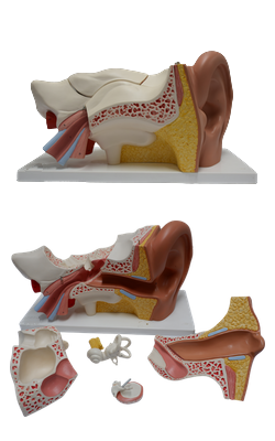
Main Model
External ear : 1 Auricle
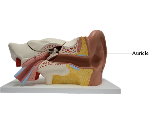
The external ear is composed of the shell-like auricle (pinna), which collects sound, and the external acoustic meatus (ear canal), which conducts sound to the tympanic membrane.
The auricle (Latin auris, ear) is composed of an irregularly shaped plate of elastic cartilage that is covered by thin skin. The auricle has several depressions and elevations. The concha of the auricle is the deepest depression. The elevated margin of the auricle is the helix. The non-cartilaginous lobule (lobe) consists of fibrous tissue, fat, and blood vessels. It is easily pierced for taking small blood samples and inserting earrings. The tragus (Greek tragos, goat; alluding to the hairs that tend to grow from this formation, like a goat’s beard) is a tongue-like projection overlapping the opening of the external acoustic meatus.
The arterial supply to the auricle is derived mainly from the posterior auricular and superficial temporal arteries.
The main nerves to the skin of the auricle are the great auricular and auriculotemporal nerves. The great auricular nerve supplies the cranial (medial) surface (commonly called the “back of the ear”) and the posterior part (helix, antihelix, and lobule) of the lateral surface (“front”). The auriculo temporal nerve, a branch of CN V3, supplies the skin of the auricle anterior to the external acoustic meatus. Minor contributions of embryological significance are made to the skin of the concha and its eminence by the vagus and facial nerves.
The lymphatic drainage of the auricle is as follows:
- the lateral surface of the superior half of the auricle drains to the superficial parotid lymph nodes;
- the cranial surface of the superior half of the auricle drains to the mastoid lymph nodes and deep cervical lymph nodes; and
- the remainder of the auricle, including the lobule, drains into the superficial cervical lymph nodes.