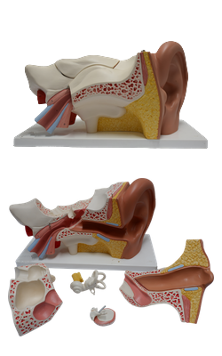
Main Model

Internal ear : 18 Cochlea

Cochlea
The cochlea is the shell-shaped part of the bony labyrinth that contains the cochlear duct, the part of the internal ear concerned with hearing. The spiral canal of the cochlea begins at the vestibule and makes 2.5 turns around a bony core, the modiolus, the cone-shaped core of spongy bone about which the spiral canal of the cochlea turns. The modiolus contains canals for blood vessels and for distribution of the branches of the cochlear nerve. The apex of the cone-shaped modiolus, like the axis of the tympanic membrane, is directed laterally, anteriorly, and inferiorly. The large basal turn of the cochlea produces the promontory of the labyrinthine wall of the tympanic cavity. At the basal turn, the bony labyrinth communicates with the subarachnoid space superior to the jugular foramen through the cochlear aqueduct. It also features the round window (Latin fenestra cochleae), closed by the secondary tympanic membrane.
Cochlear Duct
The cochlear duct is a spiral tube, closed at one end and triangular in cross section. The duct is firmly suspended across the cochlear canal between the spiral ligament on the external wall of the cochlear canal and the osseous spiral lamina of the modiolus. Spanning the spiral canal in this manner, the endolymph-filled cochlear duct divides the perilymph-filled spiral canal into two channels that are continuous at the apex of the cochlea at the helicotrema, a semilunar communication at the apex of the cochlea.
Waves of hydraulic pressure created in the perilymph of the vestibule by the vibrations of the base of the stapes ascend to the apex of the cochlea by one channel, the scala vestibuli. The pressure waves then pass through the helicotrema and descend back to the basal turn of the cochlea by the other channel, the scala tympani. Here, the pressure waves again become vibrations, this time of the secondary tympanic membrane in the round window, and the energy initially received by the (primary) tympanic membrane is finally dissipated into the air of the tympanic cavity.
The roof of the cochlear duct is formed by the vestibular membrane. The floor of the duct is also formed by part of the duct, the basilar membrane, plus the outer edge of the osseous spiral lamina. The receptor of auditory stimuli is the spiral organ (of Corti), situated on the basilar membrane. It is overlaid by the gelatinous tectorial membrane.
The spiral organ contains hair cells, the tips of which are embedded in the tectorial membrane. The organ is stimulated to respond by deformation of the cochlear duct induced by the hydraulic pressure waves in the perilymph, which ascend and descend in the surrounding scalae vestibuli and tympani.