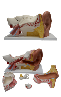
Main Model
Internal ear : 19 Vestibulocochlear nerve

Membranous Labyrinth
The membranous labyrinth consists of a series of communicating sacs and ducts that are suspended in the bony labyrinth. The labyrinth contains endolymph, a watery fluid similar in composition to intracellular fluid, thus differing in composition from the surrounding perilymph (which is like extracellular fluid) that fills the remainder of the bony labyrinth. The membranous labyrinth - composed of two divisions, the vestibular labyrinth and the cochlear labyrinth - consists of more parts than does the bony labyrinth:
• Vestibular labyrinth: utricle and saccule, two small communicating sacs in the vestibule of the bony labyrinth.
• Three semicircular ducts in the semicircular canals.
• Cochlear labyrinth: cochlear duct in the cochlea.
The spiral ligament, a spiral thickening of the periosteal lining of the cochlear canal, secures the cochlear duct to the spiral canal of the cochlea. The remainder of the membranous labyrinth is suspended by delicate filaments that traverse the perilymph.
The semicircular ducts open into the utricle through five openings, reflective of the way the surrounding semicircular canals open into the vestibule. The utricle communicates with the saccule through the utriculosaccular duct, from which the endolymphatic duct arises. The saccule is continuous with the cochlear duct through the ductus reuniens, a uniting duct. The utricle and saccule have specialized areas of sensory epithelium called maculae. The macula of the utricle (Latin macula utriculi) is in the floor of the utricle, parallel with the base of the cranium, whereas the macula of the saccule (Latin macula sacculi) is vertically placed on the medial wall of the saccule. The hair cells in the maculae are innervated by fibers of the vestibular division of the vestibulocochlear nerve. The primary sensory neurons are in the vestibular ganglia, which are in the internal acoustic meatus.
The endolymphatic duct traverses the vestibular aqueduct and emerges through the bone of the posterior cranial fossa, where it expands into a blind pouch called the endolymphatic sac. The endolymphatic sac is located under the dura mater on the posterior surface of the petrous part of the temporal bone. The sac is a storage reservoir for excess endolymph, formed by the blood capillaries in the membranous labyrinth.
Internal Acoustic Meatus
The internal acoustic meatus is a narrow canal that runs laterally for approximately 1 cm within the petrous part of the temporal bone. The internal acoustic meatus opening is in the posteromedial part of this bone, in line with the external acoustic meatus. The internal acoustic meatus is closed laterally by a thin, perforated plate of bone that separates it from the internal ear. Through this plate pass the facial nerve (CN VII), the vestibulocochlear nerve (CN VIII) and its divisions, and blood vessels. The vestibulocochlear nerve divides near the lateral end of the internal acoustic meatus into two parts: a cochlear nerve and a vestibular nerve.