Main Model
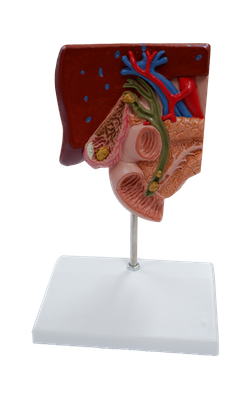
Liver
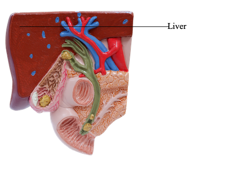
The liver is the largest gland in the body and, after the skin, the largest single organ. It weighs approximately 1500 grams and accounts for approximately 2.5% of adult body weight. In a mature fetus - when it serves as a hematopoietic organ - it is proportionately twice as large (5% of body weight).
Except for fat, all nutrients absorbed from the gastrointestinal tract are initially conveyed to the liver by the portal venous system. In addition to its many metabolic activities, the liver stores glycogen and secretes bile, a yellow-brown or green fluid that aids in the emulsification of fat.
Bile passes from the liver via the biliary ducts - right and left hepatic ducts - that join to form the common hepatic duct, which unites with the cystic duct to form the (common) bile duct. The liver produces bile continuously; however, between meals it accumulates and is stored in the gallbladder, which also concentrates the bile by absorbing water and salts. When food arrives in the duodenum, the gallbladder sends concentrated bile through the biliary ducts to the duodenum.
The liver lies mainly in the right upper quadrant of the abdomen, where it is protected by the thoracic (rib) cage and the diaphragm. The normal liver lies deep to ribs 7-11 on the right side and crosses the midline toward the left nipple. The liver occupies most of the right hypochondrium and upper epigastrium and extends into the left hypochondrium. The liver moves with the excursions of the diaphragm and is located more inferiorly when one is erect because of gravity. This mobility facilitates palpation.
The liver has a convex diaphragmatic surface (anterior, superior, and some posterior) and a relatively flat or even concave visceral surface (postero-inferior), which are separated anteriorly by its sharp inferior border that follows the right costal margin inferior to the diaphragm.
The diaphragmatic surface of the liver is smooth and dome shaped, where it is related to the concavity of the inferior surface of the diaphragm, which separates it from the pleurae, lungs, pericardium, and heart. Subphrenic recesses - superior extensions of the peritoneal cavity (greater sac) - exist between diaphragm and the anterior and superior aspects of the diaphragmatic surface of the liver. The subphrenic recesses are separated into right and left recesses by the falciform ligament, which extends between the liver and the anterior abdominal wall. The portion of the supracolic compartment of the peritoneal cavity immediately inferior to the liver is the subhepatic space.
The hepatorenal recess (hepatorenal pouch; Morison pouch) is the posterosuperior extension of the subhepatic space, lying between the right part of the visceral surface of the liver and the right kidney and suprarenal gland. The hepatorenal recess is a gravity-dependent part of the peritoneal cavity in the supine position; fluid draining from the omental bursa flows into this recess. The hepatorenal recess communicates anteriorly with the right subphrenic recess. Normally all recesses of the peritoneal cavity are potential spaces only, containing just enough peritoneal fluid to lubricate the adjacent peritoneal membranes.
The diaphragmatic surface of the liver is covered with visceral peritoneum, except posteriorly in the bare area of the liver, where it lies in direct contact with the diaphragm. The bare area is demarcated by the reflection of peritoneum from the diaphragm to it as the anterior (upper) and posterior (lower) layers of the coronary ligament. These layers meet on the right to form the right triangular ligament and diverge toward the left to enclose the triangular bare area. The anterior layer of the coronary ligament is continuous on the left with the right layer of the falciform ligament, and the posterior layer is continuous with the right layer of the lesser omentum. Near the apex (the left extremity) of the wedge-shaped liver, the anterior and posterior layers of the left part of the coronary ligament meet to form the left triangular ligament. The IVC traverses a deep groove for the vena cava within the bare area of the liver.
The visceral surface of the liver is also covered with visceral peritoneum, except in the fossa for the gallbladder and the porta hepatis - a transverse fissure where the vessels (hepatic portal vein, hepatic artery, and lymphatic vessels), the hepatic nerve plexus, and hepatic ducts that supply and drain the liver enter and leave it. In contrast to the smooth diaphragmatic surface, the visceral surface bears multiple fissures and impressions from contact with other organs.
Two sagittally oriented fissures, linked centrally by the transverse porta hepatis, form the letter "H" on the visceral surface. The right sagittal fissure is the continuous groove formed anteriorly by the fossa for the gallbladder and posteriorly by the groove for the vena cava. The umbilical (left sagittal) fissure is the continuous groove formed anteriorly by the fissure for the round ligament and posteriorly by the fissure for the ligamentum venosum. The round ligament of the liver (Latin ligamentum teres hepatis) is the fibrous remnant of the umbilical vein, which carried well-oxygenated and nutrient-rich blood from the placenta to the fetus. The round ligament and small para-umbilical veins course in the free edge of the falciform ligament. The ligamentum venosum is the fibrous remnant of the fetal ductus venosus, which shunted blood from the umbilical vein to the inferior vena cava (IVC), short- circuiting the liver.
The lesser omentum, enclosing the portal triad (bile duct, hepatic artery, and hepatic portal vein) passes from the liver to the lesser curvature of the stomach and the first 2 cm of the superior part of the duodenum. The thick, free edge of the lesser omentum extends between the porta hepatis and the duodenum (the hepatoduodenal ligament) and encloses the structures that pass through the porta hepatis. The sheet-like remainder of the lesser omentum, the hepatogastric ligament, extends between the groove for the ligamentum venosum and the lesser curvature of the stomach.
In addition to the fissures, impressions on (areas of) the visceral surface reflect the liver’s relationship to the:
• Right side of the anterior aspect of the stomach (gastric and pyloric areas).
• Superior part of the duodenum (duodenal area).
• Lesser omentum (extends into the fissure for the ligamentum venosum).
• Gallbladder (fossa for gallbladder).
• Right colic flexure and right transverse colon (colic area).
• Right kidney and suprarenal gland (renal and suprarenal areas).
Externally, the liver is divided into two anatomical lobes and two accessory lobes by the reflections of peritoneum from its surface, the fissures formed in relation to those reflections and the vessels serving the liver and the gallbladder. These superficial “lobes” are not true lobes as the term is generally used in relation to glands and are only secondarily related to the liver’s internal architecture. The essentially midline plane defined by the attachment of the falciform ligament and the left sagittal fissure separates a large right lobe from a much smaller left lobe. On the slanted visceral surface, the right and left sagittal fissures course on each side of - and the transverse porta hepatis separates - two accessory lobes (parts of the anatomic right lobe): the quadrate lobe anteriorly and inferiorly and the caudate lobe posteriorly and superiorly. The caudate lobe was so-named not because it is caudal in position (it is not) but because it often gives rise to a “tail” in the form of an elongated papillary process. A caudate process extends to the right, between the inferior vena cava (IVC) and the porta hepatis, connecting the caudate and right lobes.