Main Model
Cystic duct
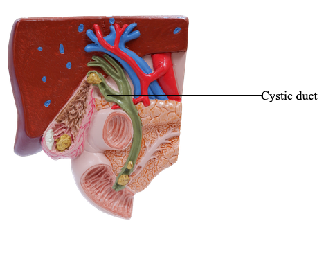
The cystic duct (3-4 cm long) connects the neck of the gallbladder to the common hepatic duct.
The cystic duct passes between the layers of the lesser omentum, usually parallel to the common hepatic duct, which it joins to form the bile duct.
The arterial supply of the cystic duct is from the cystic artery. The cystic artery commonly arises from the right hepatic artery in the triangle between the common hepatic duct, cystic duct, and visceral surface of the liver, the cystohepatic triangle (of Calot). Variations occur in the origin and course of the cystic artery.
The venous drainage from the cystic duct flows via the cystic veins. These small and usually multiple veins enter the liver directly or drain through the hepatic portal vein to the liver, after joining the veins draining the hepatic ducts and proximal bile duct.
The nerves to the cystic duct pass along the cystic artery from the celiac (nerve) plexus (sympathetic and visceral afferent [pain] fibers), the vagus nerve (parasympathetic), and the right phrenic nerve (actually somatic afferent fibers).