Main Model
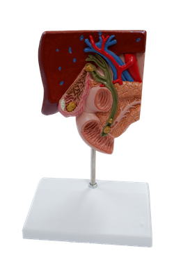
Common bile duct
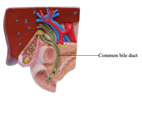
The bile duct (formerly called the common bile duct) forms in the free edge of the lesser omentum by the union of the cystic duct and common hepatic duct. The length of the bile duct varies from 5 to 15 cm, depending on where the cystic duct joins the common hepatic duct.
The bile duct descends posterior to the superior part of the duodenum and lies in a groove on the posterior surface of the head of the pancreas. On the left side of the descending part of the duodenum, the bile duct comes into contact with the main pancreatic duct. These ducts run obliquely through the wall of this part of the duodenum, where they unite, forming a dilation, the hepatopancreatic ampulla. The distal end of the ampulla opens into the duodenum through the major duodenal papilla. The circular muscle around the distal end of the bile duct is thickened to form the sphincter of the bile duct (Latin ductus choledochus). When this sphincter contracts, bile cannot enter the ampulla and the duodenum; hence, bile backs up and passes along the cystic duct to the gallbladder for concentration and storage.
The arterial supply of the bile duct is from the:
• Cystic artery: supplying the proximal part of the duct.
• Right hepatic artery: supplying the middle part of the duct.
• Posterior superior pancreaticoduodenal artery and gastroduodenal artery: supplying the retroduodenal part of the duct.
The venous drainage from the proximal part of the bile duct and the hepatic ducts usually enter the liver directly. The posterior superior pancreaticoduodenal vein drains the distal part of the bile duct and empties into the hepatic portal vein or one of its tributaries.
The lymphatic vessels from the bile duct pass to the cystic lymph nodes near the neck of the gallbladder, the lymph node of the omental foramen, and the hepatic lymph nodes. Efferent lymphatic vessels from the bile duct pass to the celiac lymph nodes.