Main Model
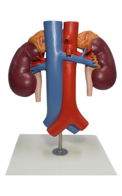
Suprarenal gland
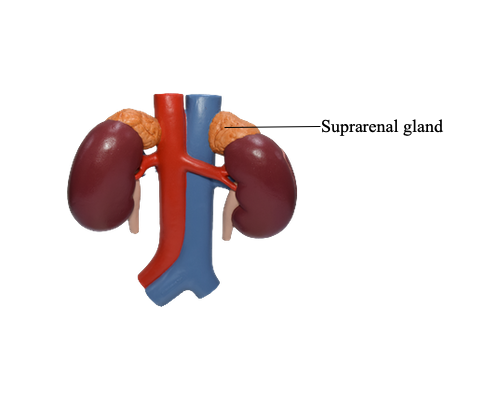
The suprarenal (adrenal) glands, yellowish in living persons, are located between the superomedial aspects of the kidneys and the diaphragm, where they are surrounded by connective tissue containing considerable perinephric fat. The suprarenal glands are enclosed by renal fascia by which they are attached to the crura of the diaphragm. Although the name “suprarenal” implies that the kidneys are their primary relationship, their major attachment is to the diaphragmatic crura. They are separated from the kidneys by a thin septum.
The shape and relations of the suprarenal glands differ on the two sides. The pyramidal right gland is more apical (situated over the superior pole) relative to the left kidney, lies anterolateral to the right crus of the diaphragm and makes contact with the inferior vena cava anteromedially and the liver anterolaterally. The crescent-shaped left gland is medial to the superior half of the left kidney and is related to the spleen, stomach, pancreas and the left crus of the diaphragm.
Each gland has a hilum, where the veins and lymphatic vessels exit the gland, whereas, the arteries and nerves enter the glands at multiple sites. The medial borders of the suprarenal glands are 4–5 cm apart. In this area, from right to left, are the inferior vena cava, right crus of the diaphragm, celiac ganglion, celiac trunk, superior mesenteric artery and the left crus of the diaphragm.
Each suprarenal gland has two parts: the suprarenal cortex and suprarenal medulla; these parts have different embryological origins and different functions.