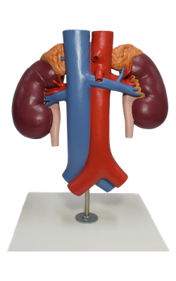
Main Model
Renal artery
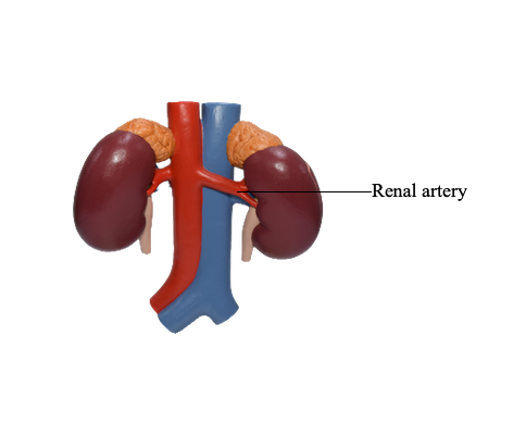
The renal arteries arise at the level of the intervertebral disc between the L1 and L2 vertebrae. The longer right renal artery passes posterior to the inferior vena cava. Typically, each artery divides close to the hilum into five segmental arteries that are end arteries (i.e., they do not anastomose significantly with other segmental arteries, so that the area supplied by each segmental artery is an independent, surgically resectable unit or renal segment). Segmental arteries are distributed to the renal segments as follows:
• The superior (apical) segment is supplied by the superior (apical) segmental artery; the anterosuperior and anteroinferior segments are supplied by the anterosuperior segmental and anteroinferior segmental arteries; and the inferior segment is supplied by the inferior segmental artery. These arteries originate from the anterior branch of the renal artery.
• The posterior segmental artery, which originates from a continuation of the posterior branch of the renal artery, supplies the posterior segment of the kidney.
Multiple renal arteries are common and usually enter the hilum of the kidney. Extrahilar renal arteries from the renal artery or aorta may enter the external surface of the kidney, commonly at their poles.