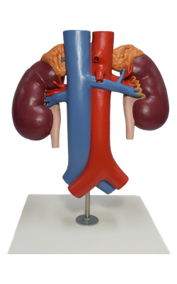
Main Model
Ureter
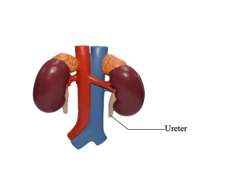
The ureters are muscular
ducts (25–30 cm long) with narrow lumina that carry urine from the
kidneys to the urinary bladder. They run inferiorly from the apices of
the renal pelves at the hila of the kidneys, passing over the pelvic
brim at the bifurcation of the common iliac arteries. They then run
along the lateral wall of the pelvis and enter the urinary bladder.
The abdominal parts of the ureters adhere closely
to the parietal peritoneum and are retroperitoneal throughout their
course. From the back, the surface marking of the ureter is a line
joining a point 5 cm lateral to the L1 spinous process and
the posterior superior iliac spine. The ureters occupy a sagittal plane
that intersects the tips of the transverse processes of the lumbar
vertebrae. When examining the ureters radiographically using contrast
medium, the ureters normally demonstrate relative constrictions in three
places:
1. At the junction of the ureters and renal pelves
2. Where the ureters cross the brim of the pelvic inlet and
3. During their passage through the wall of the urinary bladder.
These constricted areas are potential sites of
obstruction by ureteric stones (calculi). Congenital anomalies of the
ureters are fairly common.