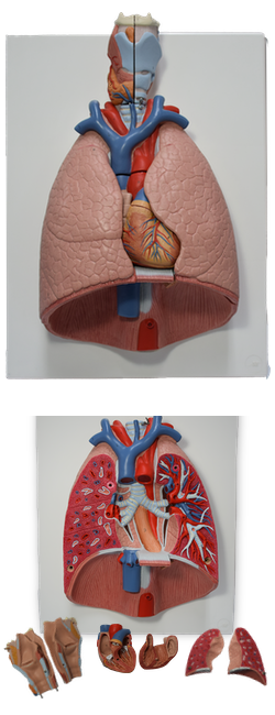
Main Model
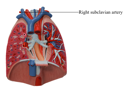
C TRACHEA : 3a Right subclavian artery
Arteries in Root of Neck
The brachiocephalic trunk is covered anteriorly by the right
sternohyoid and sternothyroid muscles; it is the largest
branch of the arch of the aorta. It arises in the midline from the beginning of the arch of the aorta, posterior to
the manubrium. It passes superolaterally to the right where
it divides into the right common carotid and right subclavian
arteries posterior to the sternoclavicular (SC) joint. The brachiocephalic trunk usually has no preterminal branches.
The subclavian arteries supply the upper limbs; they
also send branches to the neck and brain. The right subclavian artery arises from the brachiocephalic trunk. The left subclavian artery arises from
the arch of the aorta, about 1 cm distal to the left common
carotid artery. The left vagus nerve runs parallel to the first
part of the artery. Although the subclavian arteries of the two sides have different origins, their courses in
the neck begin posterior to the respective SC joints as they
ascend through the superior thoracic aperture and enter the
root of the neck.
The subclavian arteries arch superolaterally, reaching an
apex as they pass posterior to the anterior scalene muscles.
As they begin to descend, they disappear posterior to the
middle of the clavicles. As the subclavian arteries cross the
outer margin of the first ribs, their name changes; they
become the axillary arteries. Three parts of each subclavian
artery are described relative to the anterior scalene: the first
part is medial to the muscle, the second part is posterior to
it, and the third part is lateral to it.
The cervical pleurae, apices of the lung, and sympathetic
trunks lie posterior to the first part of the arteries.
The branches of the subclavian arteries are the:
• Vertebral artery, internal thoracic artery, and thyrocervical trunk from the first part of the subclavian artery.
• Costocervical trunk from the second part of the subclavian artery.
• Dorsal scapular artery, often arising from the third part of
the subclavian artery.
The cervical part of the vertebral artery arises from
the first part of the subclavian artery and ascends in the pyramidal space formed between the scalene and longus muscles
(colli and capitis). At the apex of this space, the
artery passes deeply to course through the foramina transversaria of vertebrae C1-C6. This is the vertebral part of
the vertebral artery. Occasionally, the vertebral artery may
enter a foramen more superior than vertebra C6. In approximately 5% of people, the left vertebral artery arises from the
arch of the aorta.
The suboccipital part of the vertebral artery courses
in a groove on the posterior arch of the atlas before it enters
the cranial cavity through the foramen magnum. The cranial part of the vertebral artery supplies branches to the
medulla and spinal cord, parts of the cerebellum, and the
dura of the posterior cranial fossa. At the inferior border of
the pons of the brainstem, the vertebral arteries join to form
the basilar artery, which participates in the formation of the cerebral arterial circle.
The internal thoracic artery arises from the anteroinferior aspect of the subclavian artery and passes inferomedially into the thorax. The cervical part of the internal
thoracic artery has no branches.
The thyrocervical trunk arises from the anterosuperior aspect of the first part of the subclavian artery, near the
medial border of the anterior scalene muscle. It has four
branches, the largest and most important of which is the
inferior thyroid artery, the primary visceral artery of the neck, supplying the larynx, trachea, esophagus, and thyroid
and parathyroid glands, as well as adjacent muscles. The
other branches of the thyrocervical trunk are the ascending
cervical and suprascapular arteries, and the cervicodorsal
trunk (transverse cervical artery). The terminal branches of the thyrocervical trunk are the inferior thyroid and ascending cervical arteries. The latter is a small artery that sends muscular
branches to the lateral muscles of the upper neck and spinal
branches into the intervertebral foramina.
The costocervical trunk arises from the posterior
aspect of the second part of the subclavian artery (posterior
to the anterior scalene on the right side and usually just medial to this muscle on the left side). The trunk
passes posterosuperiorly and divides into the superior intercostal and deep cervical arteries, which supply the first two
intercostal spaces and the posterior deep cervical muscles,
respectively.